. 1999 Nov;45(5):693-704.
doi: 10.1136/gut.45.5.693.

Cell specific effects of glucocorticoid treatment on the NF-kappaBp65/IkappaBalpha system in patients with Crohn's disease

Affiliations

Cell specific effects of glucocorticoid treatment on the NF-kappaBp65/IkappaBalpha system in patients with Crohn's disease

K Thiele et al. Gut. 1999 Nov.

Abstract

BACKGROUND/AIMS/PATIENTS: Glucocorticoid treatment is known to reduce nuclear factor-kappa B (NF-kappaB)p65 binding activity and activation in lamina propria cells of patients with Crohn's disease. However, lamina propria cells of glucocorticoid treated patients did not show increased expression of IkappaBalpha, and the hypothesised upregulation of IkappaBalpha by glucocorticoid treatment has not yet been shown in vivo. To investigate whether cells other than lamina propria localised mononuclear cells contribute to increased IkappaBalpha, resection gut specimens from patients matched for Crohn's disease activity index (CDAI) with or without glucocorticoid treatment were studied, and changes in the NF-kappaB/IkappaBalpha system were determined in the lamina propria as well as in underlying submucosal and endothelial cells.

Methods: Changes in the NF-kappaB/IkappaBalpha system were determined by immunohistochemistry, electrophoretic mobility shift assay, and western blot analysis in resected gut specimens from patients matched for CDAI and van Hees index with or without long term glucocorticoid treatment.

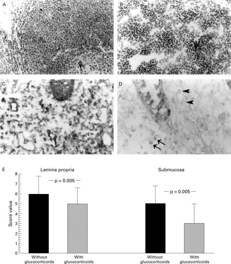
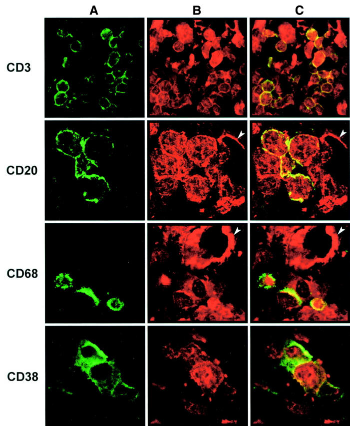
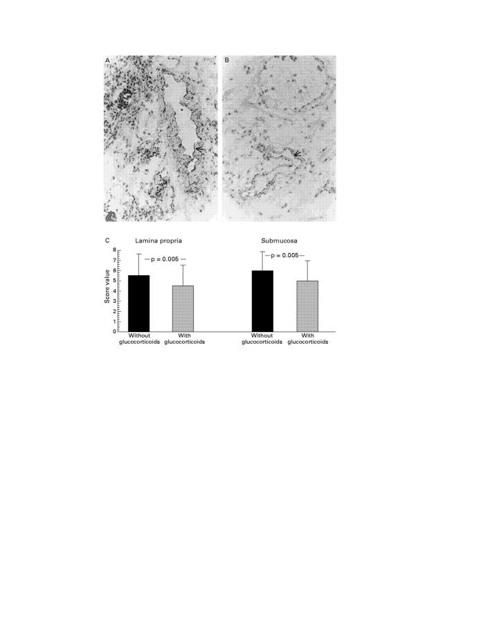
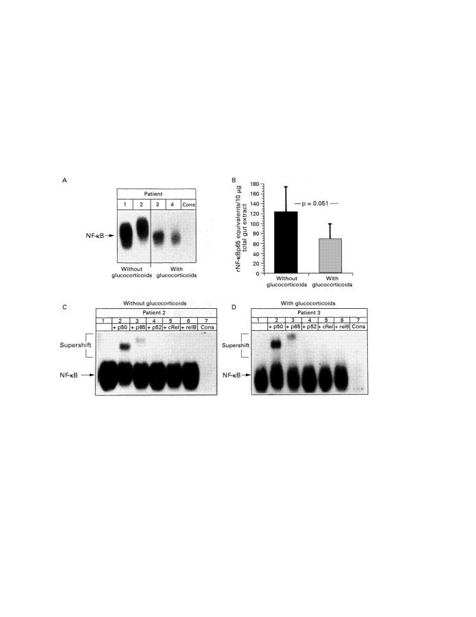
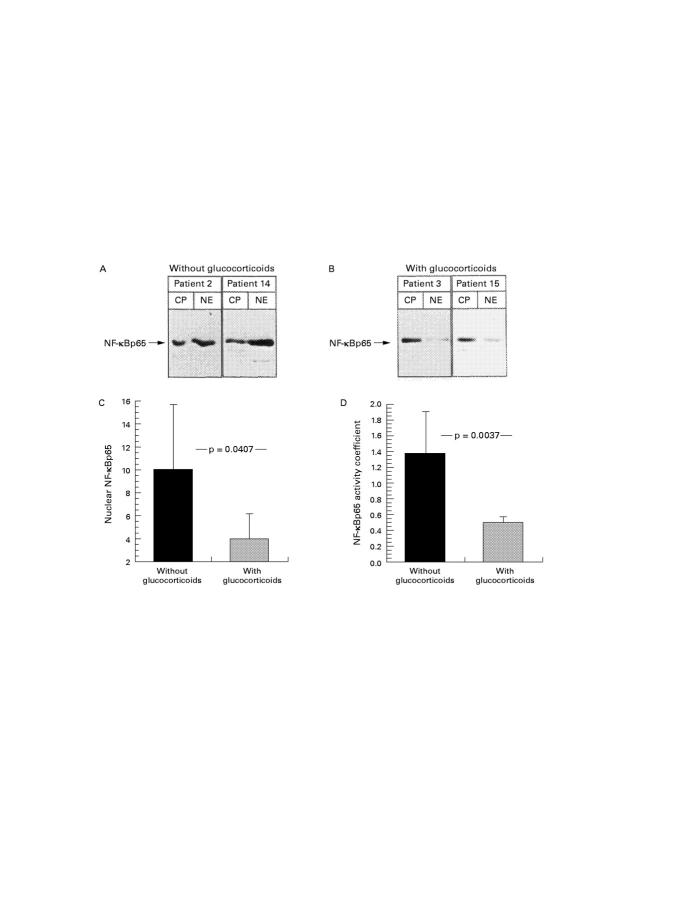
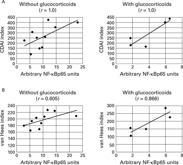
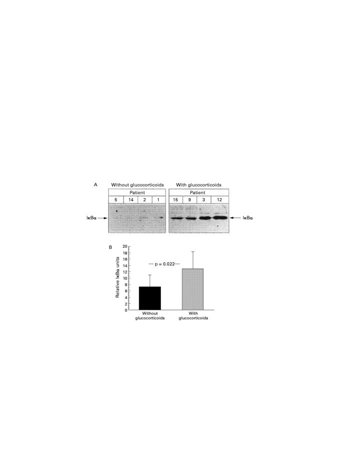
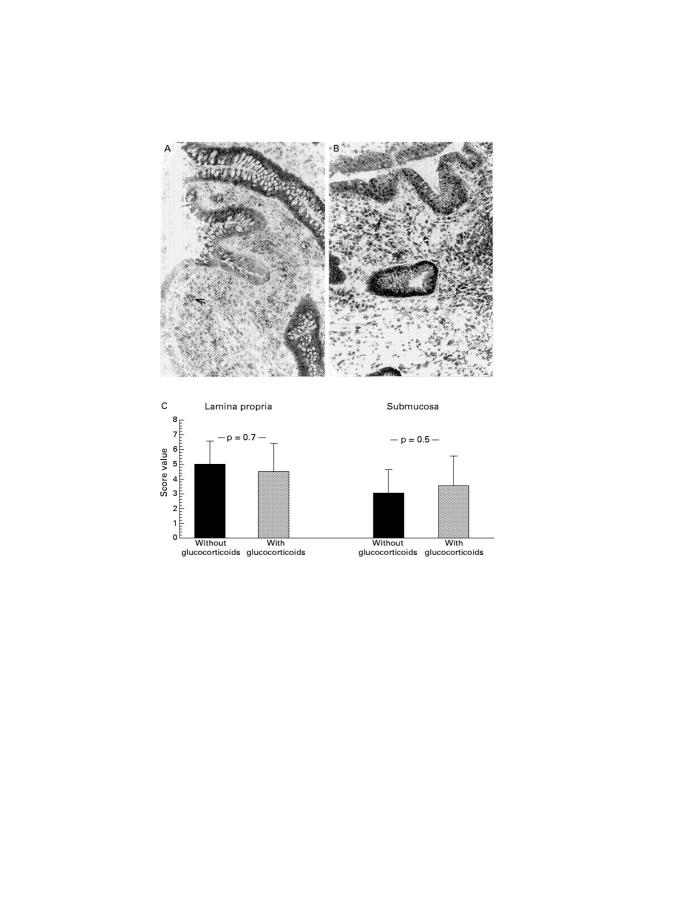
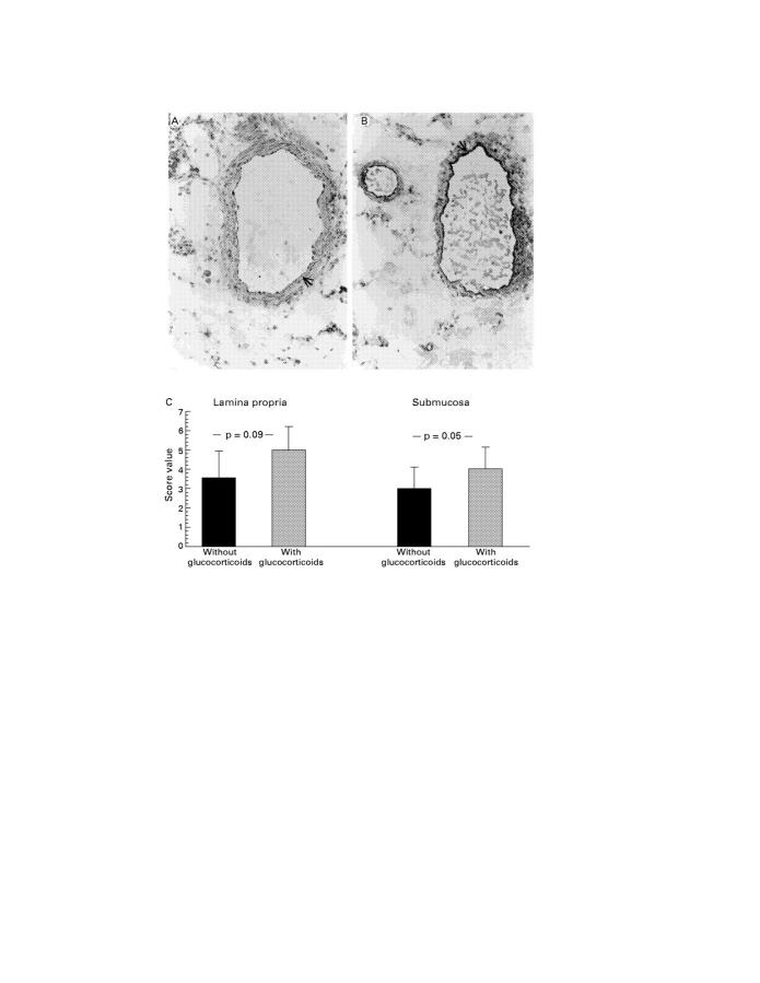
Results: Resection gut specimens from patients with Crohn's disease under glucocorticoid treatment had significantly lower nuclear NF-kappaBp65 levels in mononuclear, epithelial, and endothelial cells than samples from CDAI and van Hees index matched patients not having glucocorticoid treatment. Nuclear NF-kappaBp65 showed a strong positive correlation with both the CDAI (r = 1 for both groups) and the van Hees index (r = 0.605 for untreated and r = 0.866 for glucocorticoid treated specimens). Lower nuclear translocation of NF-kappaBp65 in the glucocorticoid treated group was paralleled by higher IkappaBalpha levels in vascular endothelial cells, but not in infiltrating mononuclear cells.

Conclusion: A comparison of resection gut specimens from untreated and treated CDAI matched patients with Crohn's disease showed downregulation of NF-kappaB binding activity and NF-kappaBp65 expression and cell specific induction of endothelial IkappaBkappa expression in the glucocorticoid treated group. As the two groups showed similar disease activity (CDAI, van Hees index), the activation of the NF-kappaBp65/IkappaBalpha system must be only part of the inflammatory cascade leading to the clinical appearance of Crohn's disease.


Figures

Figure 1
Figure 1
Immunohistochemical staining of activated NF-κBp65 antigen in resection gut specimens from patients with Crohn's disease. (A) Representative staining in a resection gut specimen from a patient without glucocorticoid treatment (original magnification × 40; representative positive stained cells are marked with an arrow). (B) Representative staining in a resection gut specimen from a patient with glucocorticoid treatment (original magnification × 40; representative positive stained cells are marked with an arrow). (C) Representative staining in a resection gut specimen from a patient without glucocorticoid treatment; counterstaining with haematoxylin was omitted (original magnification × 63). Many mononuclear cells in the lamina propria show nuclear staining. Some endothelial cells as well as crypt epithelial cells also express the molecule. (D) Representative staining in a resection gut specimen from a patient with glucocorticoid treatment; counterstaining with haematoxylin was omitted (original magnification × 100). Few mononuclear cells in the lamina propria show positive nuclear staining (arrows). Some additional positive cells display weak cytoplasmic expression of the molecule (arrowheads). (E) Summary of the scoring and the statistical results.
Figure 2
Figure 2
Double immunofluorescence staining of activated NF-κBp65 in combination with other cell markers in a resection gut specimen of a patient with Crohn's disease without glucocorticoid treatment. As leucocytes in the inflamed lamina propria show positive nuclear as well as cytoplasmic staining of NF-κBp65 (fig 1), simultaneous detection of CD3, CD20, CD68, or CD38 (column A; Cy2, green fluorescence) and NF-κBp65 (column B; Cy3, red fluorescence) was performed. This showed that NF-κBp65 is expressed by cells of all these leucocyte subsets. The double staining is shown in column C (Cy3, red; Cy2, green). Some vascular endothelial cells also express NF-κBp65 (arrowheads) (original magnifications: CD3, × 600; CD20, × 900; CD68, × 600; CD38, × 900).
Figure 3
Figure 3
Immunohistochemical staining of activated NF-κBp65 antigen in resection gut specimens from patients with Crohn's disease (original magnification × 40); representative positive stained cells are marked with an arrow. (A) Representative staining of vascular endothelial cells in a resection gut specimen from a patient without glucocorticoid treatment. (B) Representative staining of vascular endothelial cells in a resection gut specimen from a patient with glucocorticoid treatment. (C) Summary of the scoring and the statistical results.
Figure 4
Figure 4
Total gut nuclear extract (10 µg) was prepared as described in Methods and assayed for NF-κB binding activity, monitored by electrophoretic mobility shift assay (EMSA). Radioactively labelled oligonucleotides spanning the consensus NF-κB recognition motif were incubated with equal amounts of nuclear extracts, and complexes were separated by electrophoresis on non-denaturing 5% polyacrylamide gels. (A) EMSA of four representative patients without (patients 1 and 2, lanes 1 and 2) and with (patients 3 and 4, lanes 3 and 4) glucocorticoid treatment. Specificity of NF-κB binding was shown by including a 160-fold molar excess of unlabelled consensus NF-κB oligonucleotide in the reaction with the extract from patient 2 (last lane). The position of NF-κB is indicated by an arrow. (B) Nuclear extracts of all resection gut specimens (table 1) were assayed for NF-κB binding activity by EMSA. Intensity of signals was expressed as recombinant NF-κBp65 (rNF-κBp65) equivalents using an internal standard curve for rNF-κBp65 (data not shown41). (C,D) Characterisation of the NF-κB subunits contributing to the observed shift formed at the NF-κB consensus sequence was performed by including 2.5 µg of anti-p50 (lane 2), anti-p65 (lane 3), anti-p52 (lane 4), anti-cRel (lane 5), or anti-relB (lane 6) antibodies in the binding reactions with nuclear extracts of a Crohn's disease patient without (C) or with (D) glucocorticoid treatment. Specificity of NF-κB binding was shown by including a 160-fold molar excess of unlabelled consensus NF-κB oligonucleotide in the reaction with extract from patient 2 (lane 7). The position of NF-κB is indicated by arrows.
Figure 5
Figure 5
(A, B) Cytoplasmic (CP) and nuclear (NE) extracts were prepared from resection gut specimens of two patients without (patients 2 and 14; A) and two with (patients 3 and 15; B) glucocorticoid treatment as described in the Methods. Fractions were separated by SDS/PAGE (12.5% gel) and subjected to immunoblotting with an antibody for NF-κBp65. Horseradish peroxidase coupled antibodies were used as secondary antibodies, and signals were detected with the ECL-western blot detection system. The specific complexes are indicated by arrows. (C) Nuclear NF-κBp65 detected by western blot in samples without (n = 10) and with (n = 5) glucocorticoid treatment was evaluated by densitometry. The determination of the signal area to be measured and the quantitative evaluation was performed twice for two independent experiments. The mean of the two measurements was taken for statistical analysis. (D) To adjust for individual differences in NF-κB expression, the ratio of nuclear to cytoplasmic NF-κBp65 (table 4) determined in western blot analysis was calculated to give an NF-κB activity coefficient for resection gut specimens without and with glucocorticoid treatment.
Figure 6
Figure 6
Correlation of nuclear NF-κBp65 (table 4) with (A) Crohn's disease activity index and (B) van Hees index in patients without and with glucocorticoid treatment.
Figure 7
Figure 7
(A) Cytoplasmic extracts were prepared from resection gut specimens from four patients without (patient 5, 14, 2, 1; left) and four patients with (patients 15, 9, 3, 12; right) glucocorticoid treatment as described in the Methods. Fractions were separated by SDS/PAGE (12.5% gel) and subjected to immunoblotting with an antibody for full length IκBα. Horseradish peroxidase coupled antibodies were used as secondary antibodies and signals were detected with the ECL-western blot detection system. The specific complexes are indicated by arrows. (B) Cytoplasmic IκBα detected by western blot analysis in samples without (n = 10) and with (n = 5) glucocorticoid treatment was evaluated by densitometry. Determination of the signal area to be measured and the quantitative evaluation was performed twice for two independent experiments. The mean of the two measurements was taken for statistical analysis.
Figure 8
Figure 8
Immunohistochemical staining of IκBα antigen in resection gut specimens from patients with Crohn's disease (original magnification × 40); representative positive stained cells are marked with an arrow. (A) Representative staining of mononuclear cells in a resection gut specimen from a patient without glucocorticoid treatment. (B) Representative staining of mononuclear cells in a resection gut specimen from a patient with glucocorticoid treatment. (C) Summary of the scoring and the statistical results.
Figure 9
Figure 9
Immunohistochemical staining of IκBα antigen in resection gut specimens of patients with Crohn's disease (original magnification × 40); representative positive stained cells are marked with an arrow. (A) Representative staining of vascular endothelial cells in a resection gut specimen from a patient without glucocorticoid treatment. (B) Representative staining of vascular endothelial cells in a resection gut specimen from a patient with glucocorticoid treatment. (C) Summary of the scoring and the statistical results.

References

    1. Science. 1988 May 13;240(4854):889-95 - PubMed
    1. Immunobiology. 1997 Dec;198(1-3):91-8 - PubMed
    1. Cell. 1990 Apr 20;61(2):255-65 - PubMed
    1. Am J Med. 1992 Aug;93(2):199-208 - PubMed
    1. Lab Invest. 1993 May;68(5):499-508 - PubMed

Publication types

MeSH terms