. 1999 Oct 12;96(21):11866-71.
doi: 10.1073/pnas.96.21.11866.

BRCA1-associated growth arrest is RB-dependent

Affiliations
Free PMC article

BRCA1-associated growth arrest is RB-dependent

O N Aprelikova et al. Proc Natl Acad Sci U S A. .
Free PMC article

Abstract

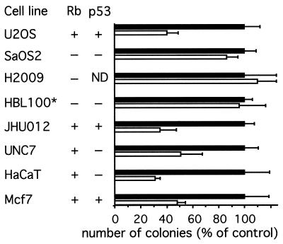
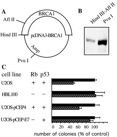
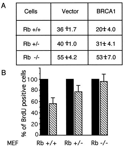
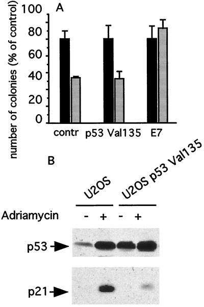
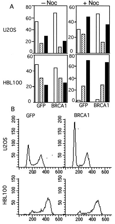
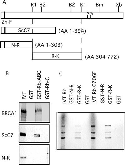
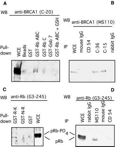
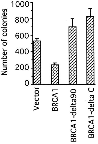
BRCA1 is a susceptibility gene for breast and ovarian cancer with growth-inhibitory activity for which the mechanism of action remains unclear. When introduced into cells, BRCA1 inhibits growth of some but not all cell lines. In an attempt to uncover the mechanism of growth suppression by BRCA1, we examined a panel of cell lines for their ability to reduce colony outgrowth in response to BRCA1 overexpression. Of all variables tested, only those cells with wild-type pRb were sensitive to BRCA1-induced growth suppression. In cells with an intact rb gene, inactivation of pRb by HPV E7 abrogates the growth arrest imposed by BRCA1. In accordance with these observations, we found that BRCA1 could not suppress BrdUrd uptake in primary fibroblasts from rb-/- mice and exhibited an intermediate ability to inhibit DNA synthesis in rb+/- as compared with rb+/+ cells. We further found that the BRCA1 protein complexes with the hypophosphorylated form of pRb. This binding is localized to amino acids 304-394 of BRCA1 protein and requires the ABC domain of pRb. In-frame deletion of BRCA1 fragment involved in interaction with pRb completely abolished the growth-suppressive property of BRCA1. Although it has been reported that BRCA1 interacts with p53, we find the p53 status did not affect the ability of BRCA1 to suppress colony formation. Our data suggest that the growth suppressor function of BRCA1 depends, at least in part, on Rb.


Figures

Figure 1
Figure 1
Growth suppression by BRCA1. Cells were transfected with equimolar amounts of pcDNA3-BRCA1 or vector alone and selected with G-418; emerging colonies were stained and counted. The number of colonies obtained with the vector alone was considered as 100%. pRb and p53 status of cells is shown. Each experiment was repeated at least three times, and error bars represent standard deviation. ■, vector alone transfections; □, BRCA1 transfections. ∗, HBL100 cell line is SV40-positive; ND, not done.
Figure 2
Figure 2
Colony formation assay was performed with the pcDNA3-BRCA1 plasmid linearized at different sites for positive and negative control. (A) Restriction map of the plasmid used in this experiment. (B) Western blot analysis of BRCA1 expression from differentially cleaved pcDNA3-BRCA1 in Cos-7 cells. (C) Colony formation assay with linearized plasmids. ■, no BRCA1 control (HindIII–AflII digest). ▧, overexpression of BRCA1 (PvuI digest).
Figure 3
Figure 3
Proliferation rate of primary mouse embyo fibroblasts transfected with BRCA1. Fibroblasts derived from rb+/+, rb−/−, or rb+/− were cotransfected with a lacZ expression vector and BRCA1 or vector alone. In 2 days, cells were labeled with BrdUrd and stained for β-gal and BrdUrd. Cells positive for β-gal were scored for proliferation assessed by BrdUrd incorporation. (A) Percentage of proliferating cells in the population of transfected cells. (B) Same as in A with the number of proliferating cells in each cell line transfected with lacZ and vector alone taken as a 100%. ■, transfection with vector alone; ▧, transfection with BRCA1. Standard deviations are presented in A and B. At least 100 β-gal-positive cells were analyzed in three fields for each transfection in two independent experiments.
Figure 4
Figure 4
BRCA1 overexpression in cells with compromised p53 or pRb. (A) Colony formation assay of U2OS control cells or U2OS overexpressing p53Val135 or E7 was performed as in Fig. 1. ■, transfection with vector alone; ░⃞, BRCA1 transfection. (B) Western blot analysis of p53 and p21 proteins induction 18 h after treatment with Adriamycin.
Figure 5
Figure 5
Overexpression of BRCA1 results in accumulation of cells in G0/G1. Cells were infected with recombinant adenovirus bearing either BRCA1 or GFP gene as control and analyzed by flow cytometry 3 days after infection. (A) Cell cycle distribution of U2OS and HBL100 cells with or without nocodazole (Noc) treatment. □, percentage of cells in G0/G1 phase of cell cycle; ░⃞, percentage of cells in S phase of cell cycle; ■, percentage of cells in G2/M phase of cell cycle. (B) DNA histogram is shown for nocodazole-treated cells described in A. At least three experiments were performed, and a representative experiment is shown.
Figure 6
Figure 6
Identification of BRCA1-Rb interactions. (A) Schematic representation of BRCA1 fragments used for in vitro translation or GST-fusion proteins. (B) In vitro-translated [35S]methionine-labeled BRCA1 fragments were incubated with GST–Rb recombinant proteins or GST alone. The complexes were collected with glutathione–Sepharose and resolved on SDS/PAGE followed by autoradiography. (C) In vitro-translated Rb or mutated Rb (C706F) proteins were used in binding to GST–BRCA1 fragments as described in B. One-fifth of the input was loaded in the IVT lane.
Figure 7
Figure 7
Rb interacts with BRCA1 protein. (A and C) Pull-down assay with the use of recombinant GST–Rb or GST–BRCA1 polypeptides. GST fusion proteins were incubated with U2OS (A) or SK-N-SH (C) cell lysates, and bound BRCA1 or Rb proteins were identified by Western blot (WB). Protein (100 μg) was loaded into WCE lanes. Approximately 1 mg of protein was used for each pull-down reaction. In A, GST-Grb7 protein was used as a negative control; in the last lane, the GST–RbABC/BRCA1 protein complex was released from the glutathione–Sepharose with glutathione (GSH) before loading as described in Methods. (B and D) Coimmunoprecipitation of pRb and BRCA1. Rb or BRCA1 proteins were immunoprecipitated from U2OS cell lysate by using anti-Rb (C-36 or C-15) (B) or anti-BRCA1 (MS110) (D). Normal mouse or rabbit IgG or anti-CD54 antibody was used as a negative control. Total protein (100 μg) was loaded into WCE lanes and ≈ 2 mg of protein was used for each immunoprecipitation.
Figure 8
Figure 8
A mutant BRCA1 protein bearing a deletion of the Rb binding domain does not suppress cell growth. Colony formation assay was performed in U2OS cells (as in Fig. 1) with wild-type BRCA1 or BRCA1 bearing an in-frame deletion of 90 aa (amino acids 303–394) or a COOH-terminal truncation. Each transfection was done in triplicate in three independent experiments.

References

    1. Easton D F, Bishop D T, Ford D, Crockford G P. Am J Hum Genet. 1993;52:678–701. - PMC - PubMed
    1. Thompson M E, Jensen R A, Obermiller P S, Page D L, Holt J T. Nat Genet. 1995;9:444–450. - PubMed
    1. Holt J T, Thompson M E, Szabo C, Robinson-Benion C, Arteaga C, King M-C, Jensen R. Nat Genet. 1996;12:298–302. - PubMed
    1. Gudas J M, Nguyen H, Li T, Cowan K H. Cancer Res. 1995;55:4561–4565. - PubMed
    1. Vaughn J P, Davis P L, Japboe M D, Huper G, Evans A C, Wiseman R W, Berchuck A, Iglehart J D, Futreal P A, Marks J R. Cell Growth Differ. 1996;7:711–715. - PubMed

Publication types

MeSH terms

Substances

LinkOut - more resources