. 1999 Oct 12;96(21):11878-82.
doi: 10.1073/pnas.96.21.11878.

Imino sugars inhibit the formation and secretion of bovine viral diarrhea virus, a pestivirus model of hepatitis C virus: implications for the development of broad spectrum anti-hepatitis virus agents

Affiliations

Imino sugars inhibit the formation and secretion of bovine viral diarrhea virus, a pestivirus model of hepatitis C virus: implications for the development of broad spectrum anti-hepatitis virus agents

N Zitzmann et al. Proc Natl Acad Sci U S A. .

Abstract

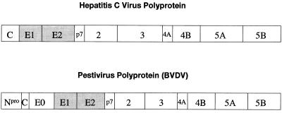
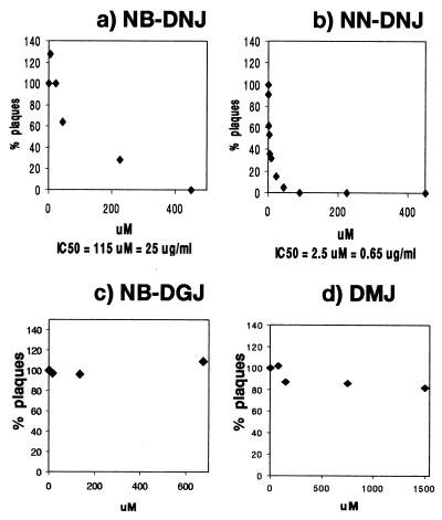
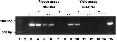
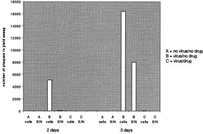
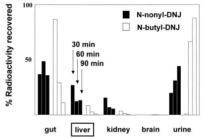
One function of N-linked glycans is to assist in the folding of glycoproteins by mediating interactions of the lectin-like chaperone proteins calnexin and calreticulin with nascent glycoproteins. These interactions can be prevented by inhibitors of the alpha-glucosidases, such as N-butyl-deoxynojirimycin (NB-DNJ) and N-nonyl-DNJ (NN-DNJ), and this causes some proteins to be misfolded and retained within the endoplasmic reticulum (ER). We have shown previously that the NN-DNJ-induced misfolding of one of the hepatitis B virus (HBV) envelope glycoproteins prevents the formation and secretion of virus in vitro and that this inhibitor alters glycosylation and reduces the viral levels in an animal model of chronic HBV infection. This led us to investigate the effect of glucosidase inhibitors on another ER-budding virus, bovine viral diarrhea virus, a tissue culture surrogate of human hepatitis C virus (HCV). Here we show that in MDBK cells alpha-glucosidase inhibitors prevented the formation and secretion of infectious bovine viral diarrhea virus. Data also are presented showing that NN-DNJ, compared with NB-DNJ, exhibits a prolonged retention in liver in vivo. Because viral secretion is selectively hypersensitive to glucosidase inhibition relative to the secretion of cellular proteins, the possibility that glucosidase inhibitors could be used as broad-based antiviral hepatitis agents is discussed. A single drug against HBV, HCV, and, possibly, HDV, which together chronically infect more than 400 million people worldwide, would be of great therapeutic value.


Figures

Figure 1
Figure 1
Polyproteins of the Flaviviridae. Viruses within the Flaviviridae (the flavi-, pesti-, and hepatitis C viruses) encode all of their proteins in a single, long ORF with the structural proteins in the N-terminal portion and the replicative nonstructural proteins in the C-terminal portion of the polyprotein (13). The polyproteins subsequently are processed by a combination of viral and host proteinases.
Figure 2
Figure 2
Secretion of infectious BVDV in the presence of N-butyl-DNJ (a), N-nonyl-DNJ (b), N-butyl-DGJ (c), and DMJ (d). (a and b) MDBK cells were grown to confluency in six-well plates and infected with 500 pfu of cp BVDV (NADL strain) per well for 1 hr at 37°. The inoculum then was replaced with medium containing the indicated concentrations of either NB-DNJ, NN-DNJ, NB-DGJ, or DMJ (plaque assay). After 3 days the supernatants were removed and used to infect fresh MDBK monolayers in six-well plates. The presence of inhibitor during the 1-hr infection does not have an effect (data not shown). After 1 hr the inoculum was removed and the cells were washed thoroughly and incubated with inhibitor-free medium. After 3 days the plaques were counted (yield assay) and the results were expressed as a percentage of the number of plaques resulting from infection with the inhibitor-free plaque assay supernatant (=100%) (x axis). The y axis indicates the inhibitor concentrations used in the plaque assay. The IC50 is indicated at the bottom. (c and d) Plaque assay results are shown. NB-DGJ was used at concentrations of up to 680 μM, an inhibitor concentration sufficient to inhibit completely the ceramide-specific glucosyltransferase (data not shown). DMJ was used at concentrations of up to 1.5 mM, an inhibitor concentration sufficient to protect treated cells from killing by a complex sugar-binding lectin (ECA) (data not shown). All experiments were done at least in duplicate, and each data point represents the average of two wells counted. Experiment 2 (b) was done in triplicate (n = 3 ± 0.13), and each data point represents the average of two wells counted.
Figure 3
Figure 3
RT-PCR of viral RNA extracted from the supernatants of NB-DNJ-treated MDBK cells. MDBK cells were grown to confluency in six-well plates, infected with cytopathic BVDV, and incubated for 3 days in the absence or presence of 4.5 mM (1,000 μg/ml) NB-DNJ. Yield assays were performed with the resulting culture medium supernatants. Viral RNA was purified from the culture medium supernatants of both plaque and yield assays, and RT-PCR was performed as described in Methods. The samples were analyzed by electrophoresis in a 1.5% agarose gel and stained with ethidium bromide. Lanes: 1, 100-bp DNA ladder; 2, culture medium from uninfected cells; 3, plaque assay culture medium from infected cells, no NB-DNJ; 4 and 5, products of a PCR on 1:10 and 1:100 dilutions, respectively, of the sample used in lane 3; 6, plaque assay culture medium from infected cells maintained in 4.5 mM NB-DNJ; 7 and 8, products of a PCR on 1:10 and 1:100 dilutions, respectively, of the sample used in lane 6; 9, yield assay culture medium from cells incubated with plaque assay supernatant from untreated cells; 10 and 11, products of a PCR on 1:10 and 1:100 dilutions, respectively, of the sample used in lane 9; 12, yield assay culture medium from cells incubated with plaque assay supernatant from NB-DNJ-treated cells; 13 and 14, products of a PCR on 1:10 and 1:100 dilutions, respectively, of the sample used in lane 12; 15, RT-PCR product using a cDNA control.
Figure 4
Figure 4
Infectivity of viral material inside and outside of NB-DNJ-treated cells 2 and 3 days after infection. Noninfected (A) and BVDV-infected (B and C) MDBK cells were grown in the absence (A and B) or presence (C) of 1 mg/ml NB-DNJ for either 2 or 3 days. The supernatants were saved and the cells were washed and lysed by freeze-thawing. Yield assays were performed to determine the number of pfus in the supernatants (S/N) and cell lysates (cells).
Figure 5
Figure 5
In vivo tissue distribution of radioactively labeled compounds. Radioactively labeled imino sugars were administered to BALB/c mice as described in Methods. After 30, 60, and 90 min, the mice were sacrificed and the organs were removed. The organs were weighed and homogenized, and aliquots were taken for radioactivity determination. Each column represents at least three animals, with cpm normalized for varying organ sizes by expressing as cpm per gram of organ homogenate.

References

    1. Alter M J. Hepatology. 1997;26, Suppl. 1:62S–65S. - PubMed
    1. Hoofnagle J H, Di Bisceglie A M. New Engl J Med. 1997;336:347–356. - PubMed
    1. Miller R H, Purcell R H. Proc Natl Acad Sci USA. 1990;87:2057–2061. - PMC - PubMed
    1. Di Bisceglie A M. Lancet. 1998;351:351–355. - PubMed
    1. Block T M, Platt F, Lu X, Gerlich W, Foster G, Blumberg B S, Dwek R A. Proc Natl Acad Sci USA. 1994;91:2235–2239. - PMC - PubMed

Publication types