. 1999 Oct 12;96(21):11895-9.
doi: 10.1073/pnas.96.21.11895.

Fgfr2 is required for limb outgrowth and lung-branching morphogenesis

Affiliations

Fgfr2 is required for limb outgrowth and lung-branching morphogenesis

E Arman et al. Proc Natl Acad Sci U S A. .

Abstract

The aim of this study was to clarify the role of Fgfr2 during later stages of embryonic development. Of two previously reported gene-targeting experiments, the more extensive Fgfr2 deletion was lethal shortly after implantation, because of trophoblast defects, whereas the less extensive one survived until midgestation with placental insufficiency and defective limb outgrowth [Xu, X., Weinstein, M., Li, C., Naski, M., Cohen, R. I., Ornitz, D. M., Leder, P. & Deng, C. (1998) Development (Cambridge, U.K.) 125, 753-765]. Fgfr2 in the early embryo is expressed in the trophectoderm, and this extra-embryonic localization persists into mid- and late gestation, when Fgfr2 also is expressed in multiple developing organs. To gain insight into the later functions of Fgfr2, fusion chimeras were constructed from homozygous mutant embryonic stem cells and wild-type tetraploid embryos. This allowed survival until term and revealed that Fgfr2 is required for both limb outgrowth and branching lung morphogenesis. The use of fusion chimeras demonstrated that early lethality was indeed because of trophectoderm defects and indicated that in the embryonic cell lineages Fgfr2 activity manifests in limb and lung development. Highly similar lung and limb phenotypes were detected recently in the loss of function mutation of Fgf10, a ligand of Fgfr2. It is likely, therefore, that whereas during early development Fgfr2 interacts with Fgf4, in limb and lung development interactions between Fgf10 and Fgfr2 may be required. Possible epithelial-mesenchymal interactions between the splicing alternatives of Fgfr2 and their specific ligands will be discussed.


Figures
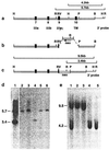
Figure 1
Figure 1
The R2Δ2 mutation of Fgfr2. (a) Genomic fragment including exons 7–10 (solid boxes). Elements of the construct are boxed, whereas the 3′ BamHI-EcoRI probe is a single line. Diagnostic HindIII and EcoRI restriction enzyme fragments recognized by the 3′ probe are shown in a and c. (b) The disruption of exon 9 (IIIc) and deletion of exon 10 (transmembrane). Arrow shows the transcriptional orientation of the neo cassette. (c) The mutant allele. The positions of the diagnostic recombinant HindIII and EcoRI fragments are shown. (d) Southern blot ES cell clones. EcoRI digest: lane 4, homozygous homologous recombinant; lane 1, heterozygous homologous recombinants; lanes 2, 3, 5, and 6, wild-type ES cell clones. (e) Southern blot of ES cell clones. HindIII digest: lanes 2 and 3, homozygous homologous recombinants; lane 1, heterozygous homologous recombinant; lanes 4 and 5, wild type. IIIa, IIIb, and IIIc, exons encoding variants of the third Ig-like loop of the ligand-binding domain; TM, transmembrane domain of Fgfr2; B, BamHI; H, HindIII; P, PstI; R, EcoRI; RV, EcoRV.
Figure 2
Figure 2
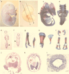
Both limb and lung development are abrogated in Fgfr2−/− tetraploid fusion chimeras. (a) 18.5 dpc. (b) 11.5 dpc. Fgfr2−/− ↔ MF1 chimeras show the absence of limbs. (c, d, and gi) Bone and cartilage preparations. Arrowheads in c show the site of the mutant scapula and pelvis. (d) Higher magnification of the pelvis in situ. Lateral (g) and dorsal (h) views of normal (Left) and mutant (Right) scapula are shown. (i) Pelvic bones (Upper, mutant; Lower, wild type). e and f show transient mesenchymal hypertrophy in the histological sections, as marked in b. (e) Hindlimb area at 11.5 dpc. (f) Forelimb area at 11.5 dpc. Sites of the hypertrophy are indicated by arrowheads. (j and k) Absence of lung development in a 14.5-dpc Fgfr2−/− ↔ MF1 chimera. (j) Wild type. (k) Mutant. (l) Cross-section of the mutant trachea displays normal histology. lu, lung; h, heart; l, liver. [Bars = 1.2 mm (b, e, and f), 1 mm (j and k), and 120 μm (l).]
Figure 3
Figure 3
Effect of the Fgfr2 mutation on the expression of Msx1 and Lhx2 in mouse embryos (9.25-dpc embryos). (A and B) Msx1 expression. (C and D) Lhx2 expression. A shows Msx1 expression in the first branchial arch and heart, the torn visceral endoderm covering the umbilical area, and the incipient forelimb bud of the wild type. Transcripts are detectable at all of these sites in the mutant (B) except the forelimb bud area, where only weak signals are seen. Lhx2 in the wild type (C) is expressed in the forebrain and facial area, the branchial arches, the heart, and the forelimb bud as well as weakly in the area of the prospective hindlimb bud. The mutant (D) is distinguished by the complete absence of Lhx2 transcripts in both limb fields. Arrows in B and D indicate the probable site of the forelimb field in the mutant.
Figure 4
Figure 4
Localized transcription of Fgfr2-IIIb, IIIc, and Fgf9. (a, d, g, and j) Bright-field illumination. (b, c, e, f, h, i, and k) Dark-field illumination. (ac) Coronal section of forelimb buds. (df) Transversal section of left hindlimb bud, 10.5 dpc. (gi) Lobes of right lung, 11.5 dpc. (j and h) Lung Fgfr2-IIIb is expressed in the surface ectoderm of the fore- and hindlimbs (b and e), as well as in the bronchial epithelium (h). Note increased Fgfr2-IIIb expression in the posterior area of the hindlimb bud (e). Fgfr2-IIIc is expressed in the mesenchyme of limb buds (c and f) and lung (i). Fgf9 is expressed in the surface ectoderm or pleura of the developing lung (k). [Bars = 150 μm (af), 200 μm (gi), and 180 μm (j and k).]

References

    1. Cohn M J, Izpisua-Belmonte J C, Abud H, Heath J K, Tickle C. Cell. 1995;80:739–746. - PubMed
    1. Crossley P H, Minowada G, MacArthur C A, Martin G R. Cell. 1996;84:127–136. - PubMed
    1. Ohuchi H, Nakagawa T, Yamamoto A, Araga A, Ohata T, Ishimaru Y, Yoshioka H, Kuwana T, Nohno T, Yamasaki M, et al. Development (Cambridge, UK) 1997;124:2235–2244. - PubMed
    1. Abud H, Skinner J A, McDonald F J, Bedford M T, Lonai P, Heath J K. Dev Genet. 1996;181:51–65. - PubMed
    1. Martin G R. Genes Dev. 1998;12:1571–1586. - PubMed

Publication types

Substances