. 1999 Oct 12;96(21):11998-2003.
doi: 10.1073/pnas.96.21.11998.

Impaired growth and fertility of cAMP-specific phosphodiesterase PDE4D-deficient mice

Affiliations

Impaired growth and fertility of cAMP-specific phosphodiesterase PDE4D-deficient mice

S L Jin et al. Proc Natl Acad Sci U S A. .

Abstract

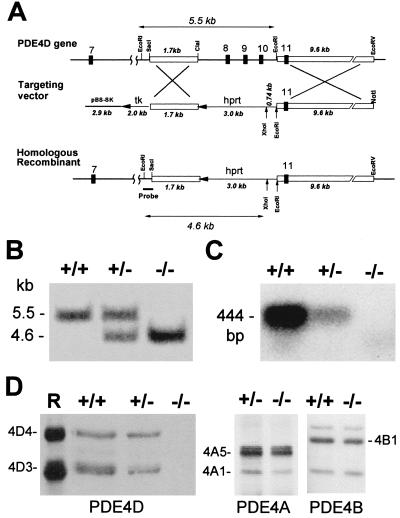
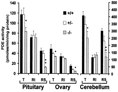
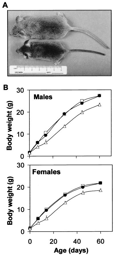
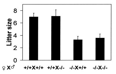
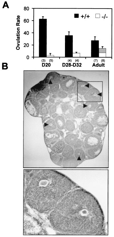
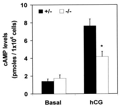
In eukaryotic cells, the inactivation of the cyclic nucleotide signal depends on a complex array of cyclic nucleotide phosphodiesterases (PDEs). Although it has been established that multiple PDE isoenzymes with distinct catalytic properties and regulations coexist in the same cell, the physiological significance of this remarkable complexity is poorly understood. To examine the role of a PDE in cAMP signaling in vivo, we have inactivated the type 4 cAMP-specific PDE (PDE4D) gene, a mammalian homologue of the Drosophila dunce. This isoenzyme is involved in feedback regulation of cAMP levels. Mice deficient in PDE4D exhibit delayed growth as well as reduced viability and female fertility. The decrease in fertility of the null female is caused by impaired ovulation and diminished sensitivity of the granulosa cells to gonadotropins. These pleiotropic phenotypes demonstrate that PDE4D plays a critical role in cAMP signaling and that the activity of this isoenzyme is required for the regulation of growth and fertility.


Figures

Figure 1
Figure 1
Targeted disruption of the mouse PDE4D locus. (A) Strategy used for targeting the PDE4D gene. The structure of the wild-type PDE4D gene in the region containing exons 7–11 (Top), the targeting vector (Middle), and the homologous recombinant (Bottom) are shown. The targeting vector derived from pBluescript SK+ (pBS-SK) contains 5′ and 3′ flanking regions of homology (open boxes) and is designed to replace the ClaI–EcoRI fragment that contains exons 8–10 with a PGK-hprt cassette. Both hprt and tk genes are in the opposite transcriptional orientation as PDE4D. The probe used for Southern blot analysis of EcoRI/XhoI-digested genomic DNA is indicated. The expected sizes of EcoRI–EcoRI (5.5 kb) and EcoRI–XhoI (4.6 kb) fragments are reported as double arrows. (B) Southern blot analysis of genomic DNA isolated from PDE4D+/+, PDE4D+/−, and PDE4D−/− mouse tail tips, digested with EcoRI and XhoI and hybridized to the probe shown in A. (C) Expression of PDE4D mRNA in mouse brain. Reverse transcription–PCR for poly(A)+ RNA extracted from pooled cortex and cerebellum of PDE4D+/+, PDE4D+/−, and PDE4D−/− mice was carried out as described in Materials and Methods. The 5′ and 3′ primers used in the PCR are specific to the exon 8 and exon 10 sequence of PDE4D, respectively. The 444-bp PCR fragment was detected by Southern blot analysis using an exon 9-specific oligonucleotide probe. (D) Expression of PDE4D, PDE4A, and PDE4B proteins in mouse brain. Cortexes and cerebella dissected from PDE4D+/+, PDE4D+/−, and PDE4D−/− mice were homogenized and immunoprecipitated as described in Materials and Methods. The PDE4D (4D3 and 4D4), PDE4A (4A1 and 4A5), and PDE4B1 proteins were detected by Western blot analysis as described in Materials and Methods. A mixture of recombinant PDE4D3 and PDE4D4 (R) was loaded for control.
Figure 2
Figure 2
PDE activity in different mouse organs. Pituitaries, ovaries, and cerebella were dissected from PDE4D+/+, PDE4D+/−, and PDE4D−/− mice and were homogenized as described in Materials and Methods. Aliquots of the homogenates were assayed for total PDE activity (T) or rolipram-insensitive (RI) PDE activity in the absence or presence of 10 μM rolipram, respectively. The rolipram-sensitive (RS) activity was obtained by subtracting the RI value from the T value. Data are the mean ± SEM (n ≥ 3 mice/genotype). Significant differences in the total activity and in the RS activity between PDE4D+/+ and PDE4D−/− mice were determined by t test and are indicated by single (*, P < 0.05) and double asterisks (**, P < 0.005).
Figure 3
Figure 3
Growth of PDE4D-deficient mice. (A) Comparison of a PDE4D−/− mouse (Lower) and a control littermate (Upper) at D34. (B) Growth curves of PDE4D+/+ (■), PDE4D+/− (●), and PDE4D−/− (▵) mice. Each point represents the mean ± SEM (n = 3–99 mice/genotype).
Figure 4
Figure 4
Female fertility of PDE4D-deficient mice. The litter size from each mating pair was determined at the day of birth. The number of mating pairs in each group is from left to right 11, 9, 13, and 17. Data are the mean ± SEM. P < 0.001, PDE4D−/− females versus wild-type females determined by ANOVA.
Figure 5
Figure 5
Ovulation of PDE4D-deficient mice. (A) Ovulation rate (oocytes/mouse) of mice at different ages of development. Wild-type (+/+) and homozygous null (−/−) mice were treated with PMSG and hCG as described in Materials and Methods. Ovulated oocytes were recovered from the ampulla of the oviducts. Data are the mean ± SEM. PDE4D−/− mice are significantly different from PDE4D+/+ mice with P < 0.001 (analyzed by ANOVA). The gray area within the bar represents the degenerated oocytes recovered in the adult mice. Numbers in the brackets indicate the number of mice used in each group. (B) Histological appearance of ovaries from pubertal PDE4D−/− mice (Upper, ×16; Lower, ×52). Mice were treated for superovulation, and ovaries were dissected, fixed, sectioned, and stained. Luteinized follicles with trapped oocytes are indicated by arrowheads.
Figure 6
Figure 6
cAMP response to hCG stimulation in granulosa cells. Granulosa cells isolated from antral follicles of the PMSG-treated mice were cultured in the absence (basal) or presence of 100 ng/ml hCG. After 1-h incubation, the intracellular cAMP accumulation was measured by RIA. Data are the mean ± SEM (n = 4 independent experiments each carried out in triplicate). The difference between PDE4D+/− and PDE4D−/− mice in response to hCG is significant (P < 0.001 determined by paired t test, *).

References

    1. Dolmetsch R E, Lewis R S, Goodnow C C, Healy J I. Nature (London) 1997;386:855–858. - PubMed
    1. Hempel C M, Vincent P, Adams S R, Tsien R Y, Selverston A I. Nature (London) 1996;384:166–169. - PubMed
    1. Beavo J A. Physiol Rev. 1995;75:725–748. - PubMed
    1. Conti M, Nemoz G, Sette C, Vicini E. Endocr Rev. 1995;16:370–389. - PubMed
    1. Lefkowitz R J. J Biol Chem. 1998;273:18677–18680. - PubMed

Publication types

Substances