

Mapping of a first locus for autosomal dominant myxomatous mitral-valve prolapse to chromosome 16p11.2-p12.1
- PMID: 10521289
- PMCID: PMC1288276
- DOI: 10.1086/302624
Mapping of a first locus for autosomal dominant myxomatous mitral-valve prolapse to chromosome 16p11.2-p12.1
Abstract
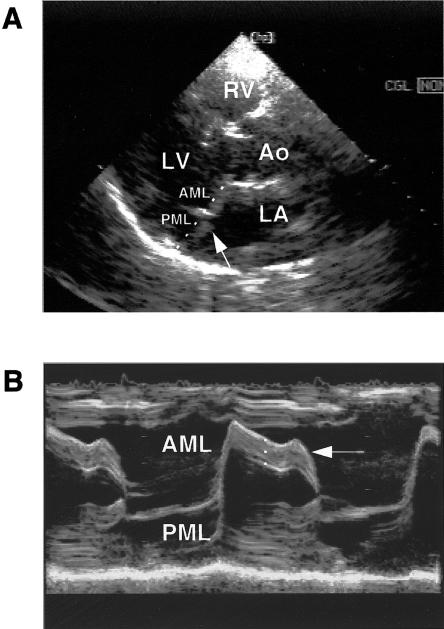
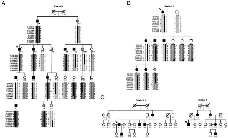
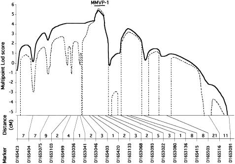
Myxomatous mitral-valve prolapse (MMVP), also called Barlow disease, is a common cardiac abnormality and affects up to 5% of the population. It is characterized by an excess of tissue that leads to billowing of the mitral leaflets, sometimes complicated by prolapse. Typical histological findings include myxomatous degeneration and degradation of collagen and elastin. Previous reports have proposed an autosomal dominant inheritance of the trait, with age- and sex-dependent expression. By systematic echocardiographic screening of the first-degree relatives of 17 patients who underwent mitral-valve repair, we have identified four pedigrees showing such an inheritance. Genomewide linkage analysis of the most informative pedigree (24 individuals, three generations) showed a significant linkage for markers mapping to chromosome 16p, with a two-point maximum LOD score for D16S3068 (Zmax=3.30 at straight theta=0). Linkage to D16S3068 was confirmed in a second family (Zmax=2.02 at straight theta=0) but was excluded for the two remaining families, thus demonstrating the genetic heterogeneity of the disease. Multipoint linkage analysis performed, with nine additional markers, on the two families with linkage gave maximum multipoint LOD scores of 5.45 and 5.68 for D16S3133, according to a conservative and a stringent model, respectively. Haplotype analysis defined a 5-cM minimal MMVP-1 locus between D16S3068 (16p11.2) and D16S420 (16p12. 1) and a 34-cM maximal interval between D16S404 and D16S3068 when recombination events were taken into account only in affected individuals. The identification of this locus represents a first step toward a new molecular classification of mitral-valve prolapse.
Figures
Comment in
-
Toward an understanding of the cause of mitral valve prolapse.Am J Hum Genet. 1999 Nov;65(5):1238-41. doi: 10.1086/302635. Am J Hum Genet. 1999. PMID: 10521288 Free PMC article. No abstract available.
References
Electronic-Database Information
-
- Généthon, http://www.genethon.fr/ (for microsatellite markers)
-
- Genome Database (GDB), http://www.gdb.org/ (for allele frequencies)
-
- Institute of Genome Research, http://www.tigr.org/ (for genomic sequences of the MMVP-1 locus)
-
- Online Mendelian Inheritance in Man (OMIM), http://www.ncbi.nlm.nih.gov/Omim (for MMVP [MIM 157700])
References
-
- Alpert JS, Sabik J, Cosgrove DM (1998) Mitral valve disease. In: Topol EJ (ed) Textbook of cardiovascular medicine. Lippincott-Raven, Philadelphia, pp 519–528
-
- Bareiss P (1976) Familial forms of the mid-end systolic click and murmur syndrome with deviations of left ventricular kinetics. Arch Mal Coeur Vaiss 69:71–81 - PubMed
-
- Barlow JB, Pocock WA (1979) Mitral valve prolapse, the specific billowing mitral leaflet syndrome, or an insignificant non-ejection systolic click. Am Heart J 97:277–285 - PubMed
-
- Barlow JB, Pocock WA, Marchand P, Denny M (1963) The significance of late systolic murmur. Am Heart J 66:443–452
-
- Boudarias JP (1991) Mitral valve prolapse: a severe abnormality? Arch Mal Coeur Vaiss 84:981–986 - PubMed
Publication types
MeSH terms
Substances
LinkOut - more resources
Full Text Sources
Medical
