. 1999 Nov;65(5):1261-7.
doi: 10.1086/302619.

The gamma-crystallins and human cataracts: a puzzle made clearer

Affiliations

The gamma-crystallins and human cataracts: a puzzle made clearer

E Héon et al. Am J Hum Genet. 1999 Nov.

Abstract

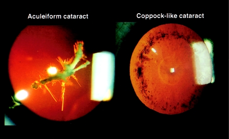
Despite the fact that cataracts constitute the leading cause of blindness worldwide, the mechanisms of lens opacification remain unclear. We recently mapped the aculeiform cataract to the gamma-crystallin locus (CRYG) on chromosome 2q33-35, and mutational analysis of the CRYG-genes cluster identified the aculeiform-cataract mutation in exon 2 of gamma-crystallin D (CRYGD). This mutation occurred in a highly conserved amino acid and could be associated with an impaired folding of CRYGD. During our study, we observed that the previously reported Coppock-like-cataract mutation, the first human cataract mutation, in the pseudogene CRYGE represented a polymorphism seen in 23% of our control population. Further analysis of the original Coppock-like-cataract family identified a missense mutation in a highly conserved segment of exon 2 of CRYGC. These mutations were not seen in a large control population. There is no direct evidence, to date, that up-regulation of a pseudogene causes cataracts. To our knowledge, these findings are the first evidence of an involvement of CRYGC and support the role of CRYGD in human cataract formation.


Figures

Figure  1
Figure 1
Clinical example of an aculeiform cataract (left) and a Coppock-like cataract (right), shown by transillumination slit-lamp photography.
Figure  2
Figure 2
Mutational analysis of CRYGC. a, Sequence chromatograms showing the single-base-pair substitution 225A→C of CRYGC exon 2, found in individuals affected with the Coppock-like–cataract phenotype. b, HphI restriction-enzyme digestion of CRYGC exon 2 of a branch of the original Coppock-like–cataract family. The wild-type fragment contains HphI restriction-enzyme sites that cut the product into fragments of 344, 82, and 358 bp, which would show one band on the agarose gel. The 225A→C mutation disrupts the second restriction-enzyme site, and products of 344 and 440 bp are observed as two bands on a stained agarose gel. Blackened and unblackened symbols represent affected and unaffected individuals, respectively. Symbols are numbered on the basis of the generation identifiers in the original publication (Lubsen et al. 1987), with all individuals numbered from left to right in ascending order.
Figure  3
Figure 3
Mutational analysis of CRYGD. a, Sequence chromatograms showing the single-base-pair substitution 411G→A of CRYGD (exon 2; nucleotides 404–418) found in individuals affected with the aculeiform-cataract phenotype. b, Mutational analysis (i.e., ARMS) of CRYGD exon 2. The ARMS assay shows cosegregation of the 411G→A mutation with the aculeiform-cataract phenotype in three Swiss families (families A–C). Selective amplification of a 194-bp PCR product for the 411G→A mutation is seen in affected individuals only. The 410-bp fragment amplified with primers for the marker D1S1663 is an internal control for the PCR reaction and is present in all lanes.
Figure  4
Figure 4
Protein-structure prediction of mutated γ-crystallin D: human γ-D crystallin prediction by SWISS-MODEL, illustrating the ARG58HIS mutation (blue) and surrounding region, protein backbone (yellow), and beta sheets (red). Note that amino acid 58 occurs between two β sheets. The R groups involved with putative hydrogen bonding are displayed; dashed green lines denote strong bonds, and dashed purple lines denote weak bonds. Left, Wild-type γ-D crystallin showing ARG58 and putative hydrogen bond between its R group (blue) and the oxygen of the carbonyl group of ILE170 near the C terminus. Right, Mutated γ-D crystallin showing HIS58 and new putative hydrogen bonds between its R group (blue) and R group of ASP171 (orange). Also predicted are hydrogen bonding with the backbone of PHE172 (at the nitrogen of the amide backbone) and weaker hydrogen bonding with the backbone of SER173 (at the nitrogen of the amide backbone).

References

Electronic-Database Information

    1. Searching GenBank, http://www.ncbi.nlm.nih.gov/Genbank/GenbankSearch.html (for sequence information on crygb and CRYGC [M19364 and K03003, respectively], CRYGD [K03005 and K03006], and CRYGE [S72943 and K03008])
    1. Online Mendelian Inheritance in Man (OMIM), http://www.ncbi.nlm.nih.gov/Omim (for aculeiform cataract [MIM 115700], Coppock-like cataract [MIM 123660 and MIM 116200], and progressive polymorphic cataract [MIM 601286])
    1. SWISS-MODEL, http://www.expasy.ch/swissmod/SWISS-MODEL.html (for protein-homology modeling)

References

    1. Brakenhoff R, Henskens H, van Rossum M, Lubsen N, Schoenmakers G (1994) Activation of the γE-crystallin pseudogene in the human hereditary Coppock-like cataract. Hum Mol Genet 3:279–283 - PubMed
    1. Cartier M, Breitman M, Tsui L-C (1992) A frame-shift mutation in the gammaE-crystallin gene of the Elo mouse. Nat Genet 2:42–45 - PubMed
    1. Cartier M, Tsui L, Ball S, Lubsen N (1994) Crystallins genes and cataract. In: Wright A, Jay B (eds) Modern genetics. Vol 2: Molecular genetics of inherited eye disorders. Harwood Academic, Edinburgh, pp 413–443
    1. Hejtmancik JF (1998) The genetics of cataract: our vision becomes clearer. Am J Hum Genet 62:520–525 - PMC - PubMed
    1. Héon E, Liu S, Billingsley G, Bernasconi O, Tsifildis C, Shorderet DF, Munier FL (1998) Gene localization for aculeiform cataract, on chromosome 2q33-35. Am J Hum Genet 63:921–926 - PMC - PubMed

Publication types