. 1999 Oct 1;13(19):2594-603.
doi: 10.1101/gad.13.19.2594.

Reconstitution of a minimal RNA degradosome demonstrates functional coordination between a 3' exonuclease and a DEAD-box RNA helicase

Affiliations

Reconstitution of a minimal RNA degradosome demonstrates functional coordination between a 3' exonuclease and a DEAD-box RNA helicase

G A Coburn et al. Genes Dev. .

Erratum in

  • Genes Dev 2000 May 1;14(9):1167

Abstract

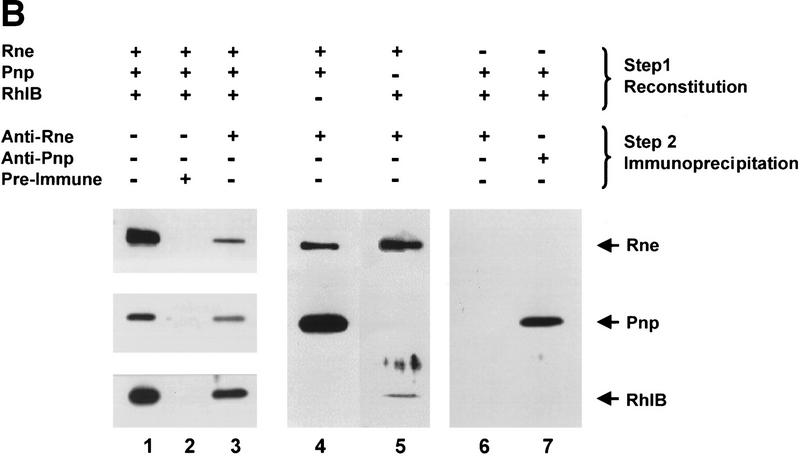
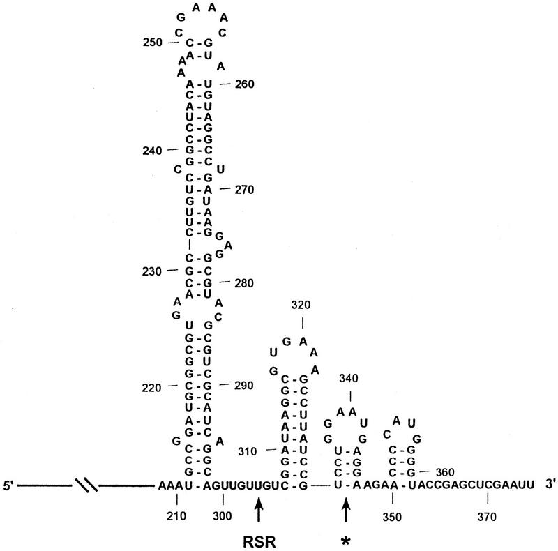
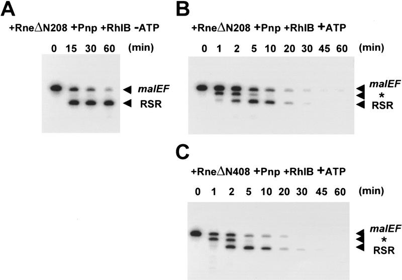
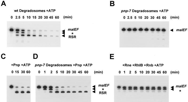
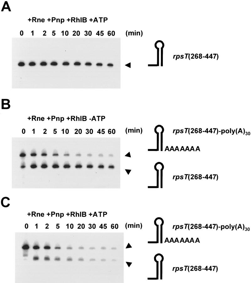
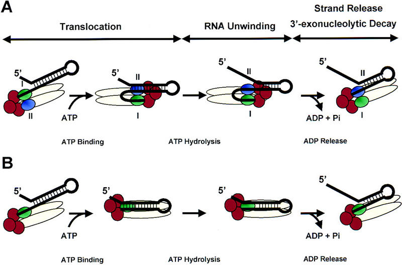
The RNA degradosome is a multiprotein complex required for the degradation of highly structured RNAs. We have developed a method for reconstituting a minimal degradosome from purified proteins. Our results demonstrate that a degradosome-like complex containing RNase E, PNPase, and RhlB can form spontaneously in vitro in the absence of all other cellular components. Moreover, ATP-dependent degradation of the malEF REP RNA by the reconstituted, minimal degradosome is indistinguishable from that of degradosomes isolated from whole cells. The Rne protein serves as an essential scaffold in the reconstitution process; however, RNase E activity is not required. Rather, Rne coordinates the activation of RhlB dependent on a 3' single-stranded extension on RNA substrates. A model for degradosome-mediated degradation of structured RNA is presented with its implications for mRNA decay in Escherichia coli.


Figures

Figure 1
Figure 1
Physical reconstitution of a minimal RNA degradosome. (A) Purified degradosome complexes and recombinant proteins used throughout this study. The following samples were denatured, separated through SDS-PAGE, and stained with Coomassie brilliant blue: (lane 1) protein molecular weight markers (Bio-Rad); (lane 2) wild-type degradosomes isolated from a wild-type strain (15 μg); (lane 3) pnp-7 degradosomes isolated from a strain containing the pnp-7 allele (12 μg); (lane 4) Rne protein (1.5 μg); (lane 5) RneΔN208 protein (1.5 μg); (lane 6) Pnp protein (1.5 μg); (lane 7) Rnb protein (1.5 μg); (lane 8) RhlB protein (1.5 μg). (B) Combinations of purified Rne, Pnp, and RhlB proteins (1 μg each) were incubated in a 40-μl volume for 20 min at 30°C. Complex formation was assayed by coimmunoprecipitation with anti-Rne, anti-Pnp, or preimmune antisera and detection by Western blotting with the appropriate antibodies as described in Materials and Methods. (Lane 1) Recombinant protein markers (100–200 ng); (lane 2) mock immunoprecipitation of the Rne/Pnp/RhlB complex with preimmune sera; (lane 3) coimmunoprecipitation of the Rne/Pnp/RhlB complex with anti-Rne antibodies; (lane 4) coimmunoprecipitation of an Rne/Pnp subcomplex with anti-Rne antibodies; (lane 5) coimmunoprecipitation of an Rne/RhlB subcomplex with anti-Rne antibodies; (lanes 6,7) a mixture of RhlB and Pnp proteins was subjected to immunoprecipitation with anti-Rne or anti-Pnp antibodies, respectively. The stoichiometry of Rne, Pnp, and RhlB in the degradosome was estimated from Coomassie blue-stained SDS–polyacrylamide gels using known amounts of each of the three components to generate standard curves. This estimation is not consistently reflected by Western analysis due to differences in the transfer efficiency of each protein, antibody titer, and exposure times.
Figure 1
Figure 1
Physical reconstitution of a minimal RNA degradosome. (A) Purified degradosome complexes and recombinant proteins used throughout this study. The following samples were denatured, separated through SDS-PAGE, and stained with Coomassie brilliant blue: (lane 1) protein molecular weight markers (Bio-Rad); (lane 2) wild-type degradosomes isolated from a wild-type strain (15 μg); (lane 3) pnp-7 degradosomes isolated from a strain containing the pnp-7 allele (12 μg); (lane 4) Rne protein (1.5 μg); (lane 5) RneΔN208 protein (1.5 μg); (lane 6) Pnp protein (1.5 μg); (lane 7) Rnb protein (1.5 μg); (lane 8) RhlB protein (1.5 μg). (B) Combinations of purified Rne, Pnp, and RhlB proteins (1 μg each) were incubated in a 40-μl volume for 20 min at 30°C. Complex formation was assayed by coimmunoprecipitation with anti-Rne, anti-Pnp, or preimmune antisera and detection by Western blotting with the appropriate antibodies as described in Materials and Methods. (Lane 1) Recombinant protein markers (100–200 ng); (lane 2) mock immunoprecipitation of the Rne/Pnp/RhlB complex with preimmune sera; (lane 3) coimmunoprecipitation of the Rne/Pnp/RhlB complex with anti-Rne antibodies; (lane 4) coimmunoprecipitation of an Rne/Pnp subcomplex with anti-Rne antibodies; (lane 5) coimmunoprecipitation of an Rne/RhlB subcomplex with anti-Rne antibodies; (lanes 6,7) a mixture of RhlB and Pnp proteins was subjected to immunoprecipitation with anti-Rne or anti-Pnp antibodies, respectively. The stoichiometry of Rne, Pnp, and RhlB in the degradosome was estimated from Coomassie blue-stained SDS–polyacrylamide gels using known amounts of each of the three components to generate standard curves. This estimation is not consistently reflected by Western analysis due to differences in the transfer efficiency of each protein, antibody titer, and exposure times.
Figure 2
Figure 2
Secondary structure of the malEF REP RNA. The 375-nucleotide malEF intragenic spacer region was folded with RNAdraw (http:///rnadraw.base8.se). The first 207 nucleotides of the malEF RNA are depicted with a line. The large REP stem–loop structure spans residues 211–298. Previously characterized intermediates generated by the stalling of exonucleases 3′ to the base of stable stem–loop structures are denoted by the asterisk (*) and RSR (McLaren et al. 1991; Py et al. 1996).
Figure 3
Figure 3
Functional reconstitution of a minimal RNA degradosome. The 375-nucleotide malEF RNA substrate (see Fig. 2) was incubated with reconstituted complexes containing recombinant Rne (750 ng/ml), Pnp (1.5 μg/ml), and/or RhlB (750 ng/ml) as indicated above each panel. As described in Materials and Methods, all incubations were performed at 30°C in the presence of 10 mm sodium phosphate and 3 mm ATP unless otherwise specified (see margins above each panel). Aliquots were removed at the times indicated (in minutes). The positions of the malEF substrate and the two major intermediates, * and RSR, are indicated with arrowheads in the margin at right of each panel. (A–C) The complex reconstituted with Rne, RhlB, and Pnp was incubated with the malEF RNA alone in the absence of ATP (A) or in the presence of ATP (B) or in the presence of 3 mm ATPγS (C). (D) The reconstituted complex was incubated with the malEF RNA in the absence of exogenous phosphate. (E–G) Subcomplexes of Rne and RhlB (E) or Rne and Pnp (F) were incubated with the malEF RNA in the presence of ATP. A mixture of RhlB and Pnp was incubated with the malEF RNA substrate in the presence of ATP (G).
Figure 4
Figure 4
RNase E activity is not required for ATP-activated degradation of the malEF RNA. The 375-nucleotide malEF RNA substrate was incubated with a reconstituted complex containing recombinant RneΔN208 (750 ng/ml), Pnp (1.5 μg/ml), and RhlB (750 ng/ml) in the absence of ATP (A) or in the presence of ATP (B). A reconstituted complex containing RneΔN408 (750 ng/ml), Pnp (1.5 μg/ml), and RhlB (750 ng/ml) was incubated with the malEF RNA in the presence of ATP (C). The products of the reaction were analyzed as described in the legend to Fig. 3. The positions of the malEF substrate and the two major intermediates, * and RSR, are indicated with arrowheads in the margin at right of each panel.
Figure 5
Figure 5
3′ exonucleases must function in cis with the Rne/RhlB RNA helicase. Degradosomes purified from a wild-type strain (wt degradosomes) or from a strain containing the pnp-7 allele of the gene encoding PNPase (pnp-7 degradosomes) were assayed in the presence of ATP against the malEF RNA substrate at 30°C as described in Materials and Methods. The products of the reaction were analyzed as described in the legend to Fig. 3. The positions of the malEF substrate and the two major intermediates, * and RSR, are indicated with arrowheads in the margin at right of each panel. (A) Kinetics of digestion of the malEF RNA by the wild-type degradosomes (5 μg/ml). (B) Kinetics of digestion of the malEF RNA by the pnp-7 degrado somes (5 μg/ml). (C) Digestion of themalEF RNA by purified PNPase (1.5 μg/ml). (D) Digestion of the malEF RNA by pnp-7 degradosomes (5 μg/ml) supplemented with recombinant PNPase (1.5 μg/ml). (E) Digestion of the malEF RNA by a partially reconstituted complex containing Rne (750 ng/ml) and RhlB (750 ng/ml) supplemented with recombinant RNase II (Rnb; 750 ng/ml).
Figure 6
Figure 6
A single-stranded 3′ end stimulates significantly the action of the minimal reconstituted degradosome. Derivatives of the rpsT mRNA were incubated with a reconstituted complex containing Rne (750 ng/ml), Pnp (575 ng/ml), and RhlB (750 ng/ml) in the presence of 3 mm ATP and 10 mm sodium phosphate as described in Materials and Methods. The products of the reaction were analyzed as described in the legend to Fig. 3. The positions of the RNA substrates and an intermediate corresponding to the removal of the poly(A)30 tail are indicated with arrowheads in the margin at right of each panel. A schematic diagram of the substrates and intermediates is also shown in the margin at right of each panel. (A) Kinetics of digestion in the absence of a poly(A) tail. The substrate, rpsT(268–447), encompasses residues 268–447 of the rpsT mRNA terminating with a flush stem–loop structure at the natural Rho-independent terminator (see text). (B) Kinetics of digestion of a polyadenylated substrate, rpsT(268–447)–poly(A)30, in the absence of ATP. The rpsT(268–447) substrate contains an additional 30 adenylate residues at its 3′ end as described in Materials and Methods. (C) Kinetics of digestion of a polyadenylated substrate in the presence of ATP. The substrate is identical to that in B.
Figure 7
Figure 7
Two potential mechanisms for RhlB-activated degradation of structured RNA by the degradosome. (A) A model prompted by the active, rolling model for DNA unwinding and translocation by the Rep DNA helicase (Wong and Lohman 1992; Korolev et al. 1997). The Rne protein (beige oval) serves as a scaffold to coordinate the action of the RhlB helicase (subunit I, green oval; subunit II, purple oval) with PNPase (red circles). This allows single-stranded RNA generated by the RNA helicase to be passed directly to the 3′ exonuclease PNPase (see text). The nucleotide-bound state of RhlB would result in a cycle of conformational and RNA affinity changes that result in translocation, RNA unwinding, and strand release/exonucleolytic degradation. (B) A second model based on the inchworm mechanism proposed for the PcrA DNA helicase (Velanker et al. 1999). In this case, RhlB acts as a monomeric protein (shown in green) that can bind both single- and double-stranded RNA. Degradation of single-stranded RNA by PNPase (red circles) takes place during the translocation step.

References

    1. Alberts B. The cell as a collection of protein machines: Preparing the next generation of molecular biologists. Cell. 1998;92:291–294. - PubMed
    1. Anderson JSJ, Parker R. The 3′ to 5′ degradation of yeast mRNA is a general mechanism for mRNA turnover that requires the SKI2 DEVH box protein and 3′ to 5′ exonucleases of the exosome complex. EMBO J. 1998;17:1497–1506. - PMC - PubMed
    1. Blum E, Py B, Carpousis AJ, Higgins CF. Polyphosphate kinase is a component of the Escherichia coli RNA degradosome. Mol Microbiol. 1997;26:387–398. - PubMed
    1. Blum E, Carpousis AJ, Higgins CF. Polyadenylation promotes degradation of 3′-structured RNA by the Escherichia coli mRNA degradosome in vitro. J Biol Chem. 1999;274:4009–4016. - PubMed
    1. Carpousis AJ, Van Houwe G, Ehretsmann C, Krisch HM. Copurification of E. coli RNase E and PNPase: Evidence for a specific association between two enzymes important for RNA processing and degradation. Cell. 1994;76:889–900. - PubMed

Publication types

MeSH terms

LinkOut - more resources