. 1999 Oct 15;520 Pt 2(Pt 2):359-71.
doi: 10.1111/j.1469-7793.1999.00359.x.

Functional expression of tagged human Na+-glucose cotransporter in Xenopus laevis oocytes

Affiliations

Functional expression of tagged human Na+-glucose cotransporter in Xenopus laevis oocytes

P Bissonnette et al. J Physiol. .

Abstract

1. High-affinity, secondary active transport of glucose in the intestine and kidney is mediated by an integral membrane protein named SGLT1 (sodium glucose cotransporter). Though basic properties of the transporter are now defined, many questions regarding the structure- function relationship of the protein, its biosynthesis and targeting remain unanswered. In order to better address these questions, we produced a functional hSGLT1 protein (from human) containing a reporter tag. 2. Six constructs, made from three tags (myc, haemaglutinin and poly-His) inserted at both the C- and N-terminal positions, were thus tested using the Xenopus oocyte expression system via electrophysiology and immunohistochemistry. Of these, only the hSGLT1 construct with the myc tag inserted at the N-terminal position proved to be of interest, all other constructs showing no or little transport activity. A systematic comparison of transport properties was therefore performed between the myc-tagged and the untagged hSGLT1 proteins. 3. On the basis of both steady-state (affinities for substrate (glucose) and inhibitor (phlorizin) as well as expression levels) and presteady-state parameters (transient currents) we conclude that the two proteins are functionally indistinguishable, at least under these criteria. Immunological detection confirmed the appropriate targeting of the tagged protein to the plasma membrane of the oocyte with the epitope located at the extracellular side. 4. The myc-tagged hSGLT1 was also successfully expressed in polarized MDCK cells. alpha-Methylglucose uptake studies on transfected cells showed an exclusively apical uptake pathway, thus indicating that the expressed protein was correctly targeted to the apical domain of the cell. 5. These comparative studies demonstrate that the myc epitope inserted at the N-terminus of hSGLT1 produces a fully functional protein while other epitopes of similar size inserted at either end of the protein inactivated the final protein.


Figures

Figure 1
Figure 1. I-V plots of glucose-induced currents for wild-type and N-terminally tagged hSGLT1
Oocytes were injected with 5 ng of either non-tagged (•) or tagged (^, myc; ▵, poly-His; □, HA) hSGLT1 cRNA. Oocytes were incubated for 5 days prior to electrophysiological assays. Glucose-induced currents were determined as described in Methods using a 5 mm glucose Barth's solution. Determination of specific currents was made by subtraction of currents measured in the absence of substrate from those in the presence of substrate. Inset, I-V plots for poly-His- and HA-tagged hSGLT1 shown on an expanded scale. Data are means ±s.e.m. of 5 oocytes from a single donor.
Figure 2
Figure 2. Determination of the affinity (Km) of hSGLT1 for glucose
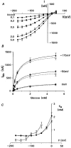
Oocytes injected with 5 ng cRNA encoding hSGLT1, myc-hSGLT1 or HA-hSGLT1 were tested for glucose-induced currents on day 5. A, glucose-specific currents from hSGLT1-injected oocytes, evaluated at increasing substrate concentration (0.1, 0.2, 0.5, 2 and 5 mm). Current in the absence of substrate was subtracted from total current in order to isolate the glucose-specific portion. B, plots of currents determined at -175, -50 and 0 mV against glucose concentration for oocytes injected with hSGLT1 (•) or myc-hSGLT1 (^) cRNA. The values were fitted according to the Michaelis-Menten equation (eqn (2)). C, affinities (Km values) for glucose determined at 25 mV increments for membrane potentials between 0 and -175 mV. Values are for oocytes injected with hSGLT1 (•), myc-hSGLT1 (^) or HA-hSGLT1 (□). Data are means ±s.e.m. of 5 oocytes from a single donor.
Figure 3
Figure 3. Determination of the affinity (Ki) of hSGLT1 for the inhibitor Pz
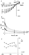
The experimental procedure for determination of the affinity for Pz was similar to that for determination of glucose affinity presented in Fig. 2. Oocytes were injected with 5 ng cRNA encoding hSGLT1 or myc-hSGLT1 and tested for Pz inhibition of glucose-induced currents after 5 days of incubation. A, I-V plots of glucose-induced currents (0.5 mm) in hSGLT1-injected oocytes in the presence of increasing amounts of Pz (0, 0.05, 0.1, 0.2, 1 and 2 μm). Non-specific current, as determined in the absence of substrate, was subtracted from total current. Data are means ±s.e.m. of 5 oocytes from a single donor. B, inhibition curves determined at two membrane potentials (-50 and -175 mV) for both hSGLT1-injected (•) and myc-hSGLT1-injected (^) oocytes. Data were plotted against Pz concentration and fitted with the equation for competitive inhibition (eqn (3)). The Km values for glucose required in the equation were determined for the same oocytes at each potential. C, Ki values for Pz determined at 25 mV steps for membrane potentials between -175 and -25 mV for oocytes injected with hSGLT1 (•) and myc-hSGLT1 (^).
Figure 4
Figure 4. Binding of Pz to oocytes
Binding of [3H]Pz was performed on control (non-injected), hSGLT1- or myc-hSGLT1-injected (50 ng cRNA) oocytes. Oocytes were incubated in groups (10 oocytes per vial) at room temperature for 15 min in Barth's solution containing the tracer. Oocytes were then rinsed 4 times with ice-cold Barth's solution and counted individually (see Methods). Data are means ±s.e.m. of counts per minute (CPM) from 8-10 oocytes from the same donor. □, binding values for specific binding as determined by subtraction of the non-specific fraction from total binding (see Methods). formula image, corresponding values of currents elicited by addition of 5 mm glucose at -175 mV.
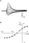
Figure 5
Figure 5. Evaluation of transient currents
A, transient current profiles determined at membrane potentials varying between -175 and +50 mV. Presteady-state currents found in normal Barth's solution were corrected for non-specific activity by subtraction of currents determined in the presence of 0.2 mm Pz. B, comparison of transient currents between oocytes expressing hSGLT1 (•) and myc-hSGLT1 (^). Oocytes were injected with 5 ng cRNA and tested on day 5. Pulse protocol and calculations of net charge transfer are described in Methods. Values are means ±s.e.m. for 3 oocytes from the same donor.
Figure 6
Figure 6. Evaluation of substrate specificity
Both hSGLT1 (□) and myc-hSGLT1 (formula image) were tested for currents induced by various sugars (Man, mannitol; 2-DG, 2-deoxyglucose; 3-OMG, 3-O-methylglucose; α-MG, α-methylglucose; and GLC, glucose. Oocytes injected with 5 ng cRNA were tested on day 5 with 5 mm substrate. Data are presented as the percentage of current found with glucose and were corrected for non-specific current by subtraction of current in the absence of sugar. Values are means ±s.e.m. of 5 oocytes from a single donor.
Figure 7
Figure 7. Western blot analysis of myc-hSGLT1 with anti-myc antibody
Plasma membrane fractions were purified from hSGLT1-injected (A) or myc-hSGLT1-injected (B) oocytes (50 ng cRNA) after 5 days of incubation. All oocytes had tested positive for glucose-induced currents of at least 1.5 μA. Samples loaded (3 μl per lane) were equivalent to 15 oocyte extracts and were shown to contain an equivalent amount of protein by Ponceau Red staining. Molecular masses indicated on the left (in kDa) were obtained using prestained molecular mass standards from New England Biolabs.
Figure 8
Figure 8. Immunofluorescence detection of myc-hSGLT1 in oocytes with anti-myc antibody
In situ identification of myc-hSGLT1 on semi-thin sections (A and B) or on untreated, intact oocytes (C and D). Oocytes were injected with hSGLT1 (A and C) or myc-hSGLT1 (B and D) and tested positive for glucose-induced currents (IGlc > 1.5 μA at -175 mV). Anti-myc antibody was used at 1:500 dilution and FITC-conjugated anti-mouse IgG at 1:250. Images were taken with a Nikon × 40 oil-immersion objective. Scale bar for A-D, 20 μm.
Figure 9
Figure 9. Immunofluorescence detection of myc-hSGLT1 in MDCK cells
MDCK cells transfected with myc-hSGLT1 (A) and non-transfected cells (B) were incubated with anti-myc antibody (1:500) and FITC-conjugated anti-mouse IgG at 1:250. Cells were seeded on coverslips at 0.35 × 10⁶ per well in 6-well plates and tested for immunofluorescence on day 4. Images were taken with a Nikon × 40 oil-immersion objective. Scale bar for A and B, 20 μm.
Figure 10
Figure 10. α-MG uptake in transfected MDCK cells
Polarized MDCK cells transfected with myc-hSGLT1 were tested for [14C]α-MG uptake activity and compared with non-transfected cells. Cells were grown on filters for 5 days after confluency and tested for both apical (□) and basolateral (formula image) uptake. The non-specific uptake, as determined by addition of 0.2 mm Pz in the uptake medium, was subtracted from total uptake measured in the absence of Pz. A 48-fold accumulation of α-MG was observed in myc-hSGLT1-transfected cells when substrate was presented on the apical side. No specific uptake of substrate was demonstrated into control cells or from the basolateral compartment of myc-hSGLT1-expressing cells.

References

    1. Bissonnette P, Gagné H, Blais A, Berteloot A. 2-Deoxyglucose transport and metabolism in Caco-2 cells. American Journal of Physiology. 1996a;270:G153–162. - PubMed
    1. Bissonnette P, Gagné H, Coady MJ, Benabdallah K, Lapointe JY, Berteloot A. Kinetic separation and characterisation of three sugar transport modes in Caco-2 cells. American Journal of Physiology. 1996b;270:G833–843. - PubMed
    1. Blais A, Bissonnette P, Berteloot A. Common characteristics for Na+-dependent sugar transport in Caco-2 cells and human fetal colon. Journal of Membrane Biology. 1987;99:113–125. - PubMed
    1. Chen X-Z, Coady MJ, Jalal F, Wallendorff B, Lapointe J-Y. Sodium leak pathway and substrate binding order in the Na+-glucose cotransporter. Biophysical Journal. 1997;73:2503–2510. - PMC - PubMed
    1. Chen X-Z, Coady MJ, Lapointe J-Y. Fast voltage clamp discloses a new component of presteady-state currents from the Na+-glucose cotransporter. Biophysical Journal. 1996;71:2544–2552. - PMC - PubMed

Publication types

MeSH terms

LinkOut - more resources