. 1999 Oct 15;520 Pt 2(Pt 2):383-92.
doi: 10.1111/j.1469-7793.1999.00383.x.

Functional MRI localisation of central nervous system regions associated with volitional inspiration in humans

Affiliations

Functional MRI localisation of central nervous system regions associated with volitional inspiration in humans

K C Evans et al. J Physiol. .

Abstract

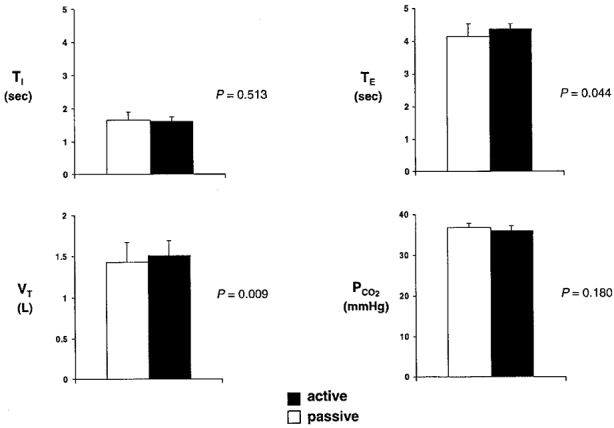
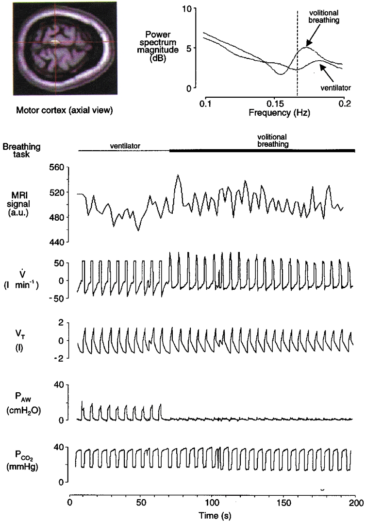
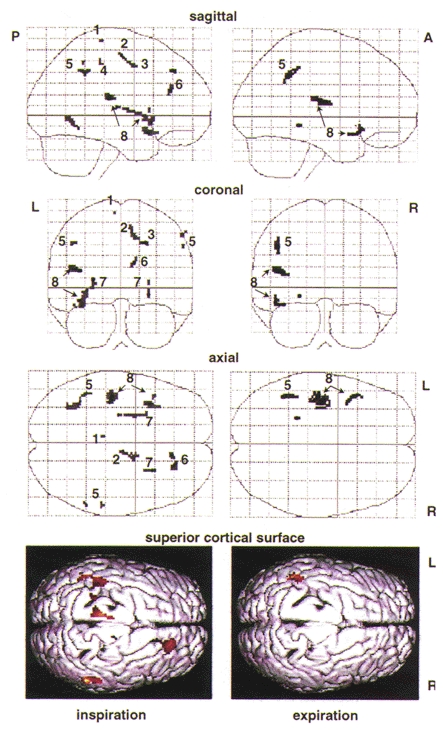
1. Functional magnetic resonance imaging (fMRI) provides a means of studying neuronal circuits that control respiratory muscles in humans with better spatial and temporal resolution than in previous positron emission tomography (PET) studies. 2. Whole brain blood oxygenation level-dependent (BOLD) changes determined by fMRI were used to identify areas of neuronal activation associated with volitional inspiration in five healthy men. Four series of scans of each subject were acquired during voluntary breathing (active task) and mechanical ventilation (passive task). Ventilation and end-tidal PCO2 were similar between tasks. Scan data were re-aligned to correct for movement artefacts and cross-referenced breath by breath to respiratory data for selective averaging of inspiratory and expiratory images. 3. Group analysis identified significant increases in the fMRI signal with volitional inspiration in the superior motor cortex, premotor cortex and supplementary motor area at loci similar to those detected in earlier studies that used PET. Additional regions activated by volitional inspiration included inferolateral sensorimotor cortex, prefrontal cortex and striatum (these foci were only revealed by PET under significant inspiratory load). 4. This study represents the first synchronised breath-by-breath analysis of respiratory-related neuronal activity with whole brain imaging in humans. Temporal resolution is sufficient to distinguish individual breaths at a normal breathing frequency.


Figures

Figure 1
Figure 1
Group mean respiratory data (n= 5) during fMRI scanning: TI, inspiratory time; TE, expiratory time; VT, tidal volume; PCO2, partial pressure of end-tidal CO2. The statistical significance between the active and passive task for each variable is represented by P (Student's paired 2-tailed t test, α = 0.05).
Figure 2
Figure 2
Record from a typical subject (F) during mechanical ventilation (passive task; first minute only) and volitional inspiration (active task; remainder of scan series). The left inset depicts an axial fMRI image and a region of interest in the left motor cortex (centre of cross-hairs). The fMRI signal (in arbitrary units, a.u.) at this region is time-aligned with the respiratory waveforms: V, airflow; VT, tidal volume; PAW, airway pressure; PCO2, partial pressure of end-tidal CO2. (NB, the ‘raw’ fMRI signal in the figure is unadjusted for haemodynamic lag.) There was a small average increase in fMRI signal during volitional inspiration relative to passive ventilation in this superior region of motor cortex. There were also phasic fMRI changes synchronised with inspiration and expiration that are particularly evident during the active task. This synchronisation (between neural activation and volitional inspiration) is depicted in the power spectra of these fMRI signals (right inset). The fMRI spectrum during volitional inspiration has an isolated peak near the frequency of breathing (0.167 Hz; vertical dashed line), whereas the spectrum during passive mechanical ventilation shows less specificity with breathing frequency.
Figure 3
Figure 3
Maximum intensity projections of multisubject analyses from selective averaging of inspiratory and of expiratory images demonstrate increases in local fMRI signal intensity during volitional inspiration (left panel, active inspiration vs. passive inspiration) and expiration (right panel, ‘passive’ expiration during the volitional breathing task vs. passive expiration during mechanical ventilation). Increases in signal intensity are represented by an arbitrary grey scale (light grey indicates statistical threshold of P= 0.001, darker shades indicate increasingly higher effect size (increasing Z values)). Plots are shown in normalised stereotactic space (see Methods) in sagittal, coronal and axial planes. A, anterior; P, posterior; R, right; L, left. Relevant local maxima are labelled: 1, superior motor cortex; 2, supplementary motor area; 3, premotor area; 4, inferolateral motor cortex; 5, inferolateral sensory cortex; 6, prefrontal cortex; 7, striatum (putamen); 8, superior temporal lobe and auditory cortex. At the bottom, a cortical surface rendering in the superior axial plane is provided for three-dimensional visualisation of significant cortical surface regions that selectively activate during volitional inspiration, left, and expiration, right (red indicates statistical threshold of P= 0.01, lighter colours indicate increasingly higher effect size (increasing Z values)).

References

    1. Bandettini PA, Jesmanowicz A, Wong EC, Hyde JS. Processing strategies for time-course data sets in functional MRI of the human brain. Magnetic Resonance in Medicine. 1993;30:161–173. - PubMed
    1. Binder JR, Rao SM, Hammeke TA, Yetkin FZ, Jesmanowicz A, Bandettini PA, Wong EC, Estkowski LD, Goldstein MD, Haughton VM, Hyde JS. Functional magnetic resonance imaging of the human auditory cortex. Annals of Neurology. 1994;35:662–672. - PubMed
    1. Boynton GM, Engel SA, Glover GH, Heeger DJ. Linear systems analysis of functional magnetic resonance imaging in the human V1. Journal of Neuroscience. 1996;16:4207–4221. - PMC - PubMed
    1. Colebatch JG, Adams L, Murphy K, Martin AJ, Lammertsma AA, Tochon-Danguy HJ, Clark JC, Friston KJ, Guz A. Regional cerebral blood flow during volitional breathing in man. The Journal of Physiology. 1991;443:91–103. - PMC - PubMed
    1. Dale AM, Buckner RL. Selective averaging of rapidly presented individual trials using fMRI. Human Brain Mapping. 1997;5:329–340. - PubMed

Publication types

LinkOut - more resources