. 1999 Oct 15;520 Pt 2(Pt 2):451-61.
doi: 10.1111/j.1469-7793.1999.t01-1-00451.x.

Nitric oxide increases persistent sodium current in rat hippocampal neurons

Affiliations

Nitric oxide increases persistent sodium current in rat hippocampal neurons

A K Hammarström et al. J Physiol. .

Abstract

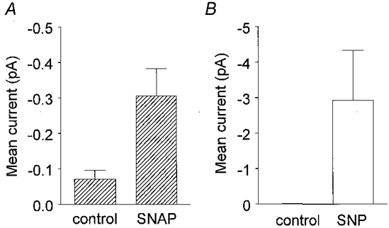
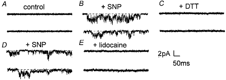
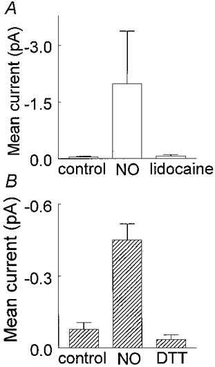
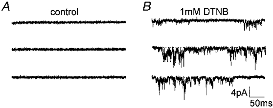
1. The effects of nitric oxide (NO) donors on whole-cell, TTX-sensitive sodium currents and single sodium channels in excised patches were examined in rat hippocampal neurons. The whole-cell sodium current consisted of a large transient component (INa,t) and a smaller, inactivation-resistant, persistent component (INa,p). 2. In acutely dissociated neurons, the amplitude of the whole-cell INa, p increased by 60-80 % within a few minutes of exposure to either of two NO donors, sodium nitroprusside (SNP, 100 microM) or S-nitroso-N-acetyl-DL-penicillamine (SNAP, 100 microM). 3. The amplitude of INa,t was not changed significantly by the same concentrations of SNP and SNAP, indicating that NO had a selective effect on INa,p. 4. Both NO donors significantly increased the mean persistent current in excised inside-out patches from cultured hippocampal neurons. SNP at 10-100 microM increased average mean persistent current at a pipette potential (Vp) of +30 mV from -0.010 +/- 0.014 pA (control) to -2.91 +/- 1.41 pA (n = 10). SNAP at 3-100 microM increased the average mean inward current in six inside-out patches from -0.07 +/- 0.02 to -0.30 +/- 0.08 pA (Vp = +30 mV). 5. The increase in persistent Na+ channel activity recorded in inside-out patches in the presence of SNP or SNAP could be reversed by the reducing agent dithiothreitol (DTT, 2-5 mM) or by lidocaine (1-10 microM). 6. The average mean current recorded in the presence of SNP was 10-fold higher than that elicited by SNAP. The time delay before an increase was observed was shorter with SNP (4.0 +/- 0.8 min, n = 8) than with SNAP (8.4 +/- 1.6 min, n = 7). 7. A component of the SNP molecule added on its own, 5 mM sodium cyanide (NaCN), increased mean current in excised inside-out patches (Vp = +30 mV) from -0.06 +/- 0.04 to -0.58 +/- 0.21 pA (n = 19). This increase in channel activity could be blocked by 10 microM lidocaine and 2-5 mM DTT. 8. These results suggest that NO may directly increase the activity of neuronal persistent Na+ channels, but not transient Na+ channels, through an oxidizing action directly on the channel protein or on a closely associated regulatory protein in the plasma membrane.


Figures

Figure 1
Figure 1. Stability of measurements of INa,p over time
The amplitude of averaged TTX-subtracted INa,p amplitudes (from 8 hippocampal neurons) measured at the end of 400 ms depolarizing pulses relative to the amplitude of INa,p recorded when first obtaining whole-cell seal (Normalised current) plotted against time from the beginning of the whole-cell recording. There was no significant change in the amplitude for more than 15 min of recording.
Figure 2
Figure 2. Effect of NO donors on whole-cell Na+ currents in CA1 neurons
Currents were generated by a depolarising step to -30 mV (A) or -20 mV (B and C) from a holding potential of -100 mV. The traces shown are TTX subtracted (see Methods). A, currents recorded from a neuron before (upper trace) and after exposure to 0.1 mm SNP for 7.5 min and after 12 min of wash-out (lower traces). In this neuron, 12 min of wash-out did not reverse the effect of SNP. B, TTX-sensitive currents recorded from a neuron before (upper trace) and after 3 and 10 min of exposure to 0.1 mm SNAP (lower traces). An increase in the amplitude of INa,p was seen after 3 min and the amplitude of INa,p was doubled after 10 min of 0.1 mm SNAP perfusion. C, same neuron as in B but at a lower vertical amplification to show the lack of comparable effect of 0.1 mm SNAP on INa,t.
Figure 3
Figure 3. Current-voltage and conductance-voltage relationships for INa,p before and after 0.1 mm SNAP
A, current-voltage relationship of the TTX-sensitive persistent Na+ current before (^) and after a 10 min 0.1 mm SNAP exposure (•) (same cell as in Fig. 2B and C). B, conductance-voltage relationship of the TTX-sensitive persistent Na+ current before (^) and after 10 min of 0.1 mm SNAP (•) (same cell as in A). The amplitude of INa,p was divided by V - V0 to obtain the conductance (V0 is the potential at which I= 0). The lines through the data points were the best fits of the equation G = Gmax/(1 + exp((V’ - V)/k)). The line of best fit gave a Gmax of 1.5 nS before SNAP exposure and 2.4 nS in the presence of SNAP. V’ and slope factors were -41 and 12 mV in control and -42 and 13 mV in the presence of SNAP. Further details are given in the text.
Figure 5
Figure 5. Increases in INa,p produced by SNP and SNAP in inside-out patches
The histograms show mean current amplitudes. The vertical lines denote ± 1 s.e.m.A, mean data from 6 patches before (control, -0.07 ± 0.02 pA) and after 3-100 μm SNAP (-0.30 ± 0.08 pA, P= 0.02). B, mean data from 10 patches before (control, -0.010 ± 0.014 pA, n= 10) and after 10-100 μm SNP (-2.91 ± 1.41 pA, n= 10, P= 0.05).
Figure 4
Figure 4. Effect of SNAP on INa,p activity in an excised inside-out patch
The representative 400 ms traces shown were obtained from 3-4 min recordings from a patch held at Vp=+30 mV. The accompanying all-points histograms were calculated from the full 3-4 min current record sampled at 10 kHz. A, control activity. B, increased activity after 5 min exposure to 0.1 mm SNAP. C, SNAP-induced activity was completely blocked after 4-5 min exposure to 10 μm lidocaine and 0.1 mm SNAP.
Figure 6
Figure 6. Effects of SNP, DTT and lidocaine on an inside-out patch
Current traces were obtained at a holding potential of -30 mV (Vp=+30 mV) and are all from the same patch. A, control current records before exposure to SNP. B, after approximately 4 min in the presence of 10 μm SNP. C, inhibition of channel activity after 2-3 min of perfusion with 2 mm DTT (with 10 μm SNP). D, the return of activity after 1.5 min of washing out DTT with a solution containing 10 μm SNP. E, abolition of SNP-elicited channel activity by 10 μm lidocaine (with 10 μm SNP, 10 min).
Figure 7
Figure 7. Average effects of DTT and lidocaine on current activity produced by SNP or SNAP
Histograms show averaged data and the vertical bars denote 1 s.e.m.A, mean current was measured from 5 patches before (-0.04 ± 0.02 pA), and after exposure to 3-100 μm SNP or SNAP (≈5.5 min exposure, -2.46 ± 1.70 pA) and then in the presence of 10 μm lidocaine plus NO donor (-0.07 ± 0.04 pA). B, mean current was measured in 4 excised patches before (-0.03 ± 0.02 pA), and after approximately 7.5 min exposure to SNP or SNAP (-0.45 ± 0.07 pA) and then after 2-3 min perfusion with 2-5 mm DTT plus NO donor (-0.03 ± 0.04 pA).
Figure 8
Figure 8. Effect of NaCN on Na+ channel activity
Representative traces are from an inside-out patch held at Vp=+30 mV. A, traces before drug application. B, after a 5 min application of 5 mm NaCN. C, inhibition of activity by 10 μm lidocaine.
Figure 9
Figure 9. The effect of 5 mm NaCN on mean persistent Na+ current
Mean current recorded from 19 inside-out patches in control (-0.06 ± 0.04 pA), after approximately 5 min of exposure to 5 mm NaCN (-0.58 ± 0.21 pA) and after lidocaine exposure (-0.01 ± 0.01 pA, n= 4). NaCN significantly increased inward persistent channel activity which could be abolished by 10 μm lidocaine. The difference between control and 5 mm NaCN is statistically significant (n= 19, P= 0.02) as is the difference between 5 mm NaCN and 10 μm lidocaine (n= 4, P= 0.01).
Figure 10
Figure 10. Effect of 1 mm DTNB on inside-out hippocampal patch
A sulfhydryl specific reagent, DTNB, caused an increase in persistent Na+ channel activity in a patch showing no channel activity over a previous 4 min recording period. A, before 1 mm DTNB was perfused. B, after about 6 min of exposure to 1 mm DTNB.

References

    1. Alonso A, Llinás RR. Subthreshold Na+-dependent theta-like rythmicity in stellate cells of entorhinal cortex layer II. Nature. 1989;342:1175–1177. - PubMed
    1. Alzheimer C, Schwindt PC, Crill WE. Modal gating of Na+ channels as a mechanism of persistent Na+ current in pyramidal neurons from rat and cat sensorimotor cortex. Journal of Neuroscience. 1993;13:660–673. - PMC - PubMed
    1. Amitai Y. Membrane potential oscillations underlying firing patterns in neocortical neurons. Neuroscience. 1994;63:151–161. - PubMed
    1. Archer SL, Huang JM, Hampl V, Nelson DP, Shultz PJ, Weir EK. Nitric oxide and cGMP cause vasorelaxation by activation of a charybdotoxin-sensitive K channel by cGMP-dependent protein kinase. Proceedings of the National Academy of Sciences of the USA. 1994;91:7583–7587. - PMC - PubMed
    1. Arden SR, Sinor JD, Potthoff WK, Aizenman E. Subunit-specific interactions of cyanide with the N-methyl-D-aspartate receptor. Journal of Biological Chemistry. 1998;273:21505–21511. - PubMed

Publication types

LinkOut - more resources