Comparative Study
. 1999 Nov;37(11):3701-4.
doi: 10.1128/JCM.37.11.3701-3704.1999.

Similar signature of the prion protein in natural sheep scrapie and bovine spongiform encephalopathy-linked diseases

Affiliations
Comparative Study

Similar signature of the prion protein in natural sheep scrapie and bovine spongiform encephalopathy-linked diseases

T G Baron et al. J Clin Microbiol. 1999 Nov.

Abstract

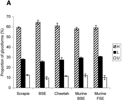
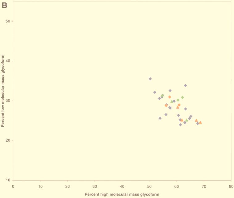
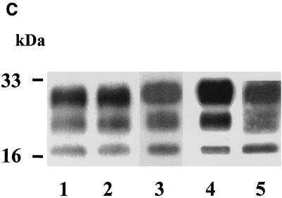
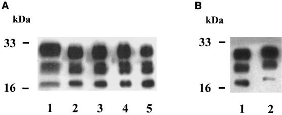
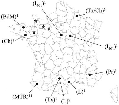
It has been suggested that specific molecular features could characterize the protease-resistant prion protein (PrP res) detected in animal species as well as in humans infected by the infectious agent strain that causes bovine spongiform encephalopathy (BSE). Studies of glycoform patterns in such diseases in French cattle and cheetahs, as well as in mice infected by isolates from both species, revealed this characteristic molecular signature. Similar studies of 42 French isolates of natural scrapie, from 21 different flocks in different regions of France, however, showed levels of the three glycoforms comparable to those found in BSE-linked diseases. Moreover, the apparent molecular size of the unglycosylated form was also indistinguishable among all different sheep isolates, as well as isolates from BSE in cattle. Overall results suggest that scrapie cases with features similar to those of BSE could be found more frequently in sheep than previously described.


Figures

FIG. 1
FIG. 1
(A) Proportions of PrP res glycoforms in transmissible SE from sheep (n = 42), cattle (n = 4), and cheetahs (n = 2) and from mice infected with BSE (n = 3) or transmissible SE from cheetahs (n = 3). Percentages are indicated as means ± standard errors of the means of the PrP res bands, H, L, and U. FSE, feline SE. (B) Scattergraph of proportions of H and L glycoforms of PrP res. Red diamonds, cattle BSE isolate-infected mice; green diamonds, cheetah SE isolate-infected mice; blue diamonds, scrapie sheep; red triangles, cattle BSE; green triangles, cheetah SE. (C) Western blot of PrP res. Lane 1, cattle BSE isolate-infected mice; lane 2, cheetah SE isolate-infected mice; lane 3, scrapie sheep; lane 4, cattle BSE; lane 5, cheetah SE.
FIG. 1
FIG. 1
(A) Proportions of PrP res glycoforms in transmissible SE from sheep (n = 42), cattle (n = 4), and cheetahs (n = 2) and from mice infected with BSE (n = 3) or transmissible SE from cheetahs (n = 3). Percentages are indicated as means ± standard errors of the means of the PrP res bands, H, L, and U. FSE, feline SE. (B) Scattergraph of proportions of H and L glycoforms of PrP res. Red diamonds, cattle BSE isolate-infected mice; green diamonds, cheetah SE isolate-infected mice; blue diamonds, scrapie sheep; red triangles, cattle BSE; green triangles, cheetah SE. (C) Western blot of PrP res. Lane 1, cattle BSE isolate-infected mice; lane 2, cheetah SE isolate-infected mice; lane 3, scrapie sheep; lane 4, cattle BSE; lane 5, cheetah SE.
FIG. 2
FIG. 2
Western blot of PrP res. (A) Lane 1, cattle BSE; lanes 2 to 5, natural sheep scrapie cases. (B) Lane 1, cattle BSE isolate-infected mouse; lane 2, scrapie-infected mouse (C506 M3 strain).
FIG. 3
FIG. 3
Geographic distribution (spots), sheep breeds, and numbers of outbreaks studied (superscript numbers) of natural scrapie (n = 21). Comparative locations of BSE cases in cattle (n = 4) are indicated (stars). Sheep breeds were Manech tête rousse (MTR), Lacaune (L), Bleu du Maine (BdM), Préalpes (Pr), Texel (Tx), Charollais (Ch), and INRA 401 (I401).

References

    1. Baron T, Belli P, Madec J-Y, Moutou F, Vitaud C, Savey M. Spongiform encephalopathy in an imported cheetah in France. Vet Rec. 1997;141:270–271. - PubMed
    1. Bolton D, Seligman S, Bablanian G, Windsor D, Scala L J, Kim K S, Chen C J, Kascsak R J, Bendheim P E. Molecular location of a species-specific epitope on the hamster scrapie agent protein. J Virol. 1991;65:3667–3675. - PMC - PubMed
    1. Bruce M E, Will R G, Ironside J W, McConnell I, Drummond D, Suttle A, McCardle L, Chree A, Hope J, Birkett C, Cousens S, Fraser H, Bostock C J. Transmissions to mice indicate that “new variant” CJD is caused by the BSE agent. Nature. 1997;389:498–501. - PubMed
    1. Collinge J, Sidle K C L, Meads J, Ironside J, Hill A F. Molecular analysis of prion strain variation and the aetiology of 'new variant' CJD. Nature. 1996;383:685–690. - PubMed
    1. Deslys J-P, Lasmézas C, Streichenberger N, Hill A, Collinge J, Dormont D, Kopp N. New variant Creutzfeldt-Jakob disease in France. Lancet. 1997;349:30–31. - PubMed

Publication types

MeSH terms

LinkOut - more resources