. 1999 Oct 18;190(8):1103-14.
doi: 10.1084/jem.190.8.1103.

Herpes simplex virus type 1 infection of activated cytotoxic T cells: Induction of fratricide as a mechanism of viral immune evasion

Affiliations

Herpes simplex virus type 1 infection of activated cytotoxic T cells: Induction of fratricide as a mechanism of viral immune evasion

M J Raftery et al. J Exp Med. .

Abstract

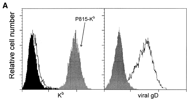
Herpes simplex virus type 1 (HSV1), a large DNA-containing virus, is endemic in all human populations investigated. After infection of mucocutaneous surfaces, HSV1 establishes a latent infection in nerve cells. Recently, it was demonstrated that HSV1 can also infect activated T lymphocytes. However, the consequences of T cell infection for viral pathogenesis and immunity are unknown. We have observed that in contrast to the situation in human fibroblasts, in human T cell lines antigen presentation by major histocompatibility complex class I molecules is not blocked after HSV1 infection. Moreover, HSV1 infection of T cells results in rapid elimination of antiviral T cells by fratricide. To dissect the underlying molecular events, we used a transgenic mouse model of HSV1 infection to demonstrate that CD95 (Apo-1, Fas)-triggered apoptosis is essential for HSV1-induced fratricide, whereas tumor necrosis factor (TNF) also contributes to this phenomenon but to a lesser extent. By contrast, neither TRAIL (TNF-related apoptosis-inducing ligand) nor perforin were involved. Finally, we defined two mechanisms associated with HSV1-associated fratricide of antiviral T cells: (a) T cell receptor-mediated upregulation of CD95 ligand and (b) a viral "competence-to-die" signal that renders activated T lymphocytes susceptible to CD95 signaling. We propose that induction of fratricide is an important immune evasion mechanism of HSV1, helping the virus to persist in the host organism throughout its lifetime.


Figures

Figure 1
Figure 1
Levels of virus-encoded Kb and endogenous MHC class I molecules on human fibroblasts and CD8+ T cells infected with HSV1. The human fibroblast cell line MRC-5 (left) and human Con A–activated PBMCs (right) were infected with Kb-encoding HSV1 strain F-US5MHC at an MOI of 10 (unfilled curve), HSV-1 strain F at an MOI of 10 (black curve), or mock infected (gray curve). After incubation for 5 h, cells were stained for viral Kb, endogenous MHC class I molecules, and viral gD using a primary mAb followed by an isotype-specific FITC-labeled secondary reagent. PBMCs were subsequently stained with a PE-conjugated anti-CD8 mAb, and data were electronically gated for CD8+ cells. The x-axis shows fluorescence intensity (log scale, four decades), and the y-axis depicts the relative cell number. The results shown are representative of three independent experiments.
Figure 2
Figure 2
Fratricide of HSV1-reactive human and murine T cell lines. HSV1-reactive T cell lines from mice and human donors were isolated as described in Materials and Methods. The expanded T cells were infected with HSV1 strain F at an MOI of 10 and analyzed in a JAM assay 5 h after infection to measure the degree of cell death. As controls, polyclonally activated human and murine T cells and HCMV-reactive T cell lines were treated in the same way. One of three independent experiments is depicted.
Figure 3
Figure 3
CTLs from Des.TCR mice recognize viral Kb as a target structure. (A) FACS™ analysis of Kb expression on target cells. P815 cells were infected with either F-US5MHC (unfilled curve) or F-US5β (black curve) at an MOI of 10 or mock infected (gray curve). After incubation for 5 h, the cells were stained for Kb (left) or gD (right). As a positive control, uninfected P815-Kb transfectants strongly expressing Kb (gray curve marked P815-Kb) were stained for Kb. The x-axis shows fluorescence intensity (log scale, three decades), and the y-axis depicts the relative cell number. (B) In parallel, target cells were used in a 51Cr-release assay. The x-axis shows the E/T ratio, whereas the y-axis depicts specific lysis (%). Results are representative of three independent experiments.
Figure 3
Figure 3
CTLs from Des.TCR mice recognize viral Kb as a target structure. (A) FACS™ analysis of Kb expression on target cells. P815 cells were infected with either F-US5MHC (unfilled curve) or F-US5β (black curve) at an MOI of 10 or mock infected (gray curve). After incubation for 5 h, the cells were stained for Kb (left) or gD (right). As a positive control, uninfected P815-Kb transfectants strongly expressing Kb (gray curve marked P815-Kb) were stained for Kb. The x-axis shows fluorescence intensity (log scale, three decades), and the y-axis depicts the relative cell number. (B) In parallel, target cells were used in a 51Cr-release assay. The x-axis shows the E/T ratio, whereas the y-axis depicts specific lysis (%). Results are representative of three independent experiments.
Figure 4
Figure 4
F-US5MHC infection and fratricide of Des.TCR cells. (A) Levels of viral Kb and glycoprotein gD on activated murine T cells infected with F-US5MHC (unfilled curve). As a control, cells were infected with F-US5β (black curve) or mock infected (gray curve). After incubation for 5 h, the cells were stained for viral Kb and gD using a primary mAb followed by an isotype-specific FITC-labeled secondary reagent. Splenocytes were subsequently stained with PE-conjugated anti-CD8 mAb, and data were electronically gated for CD8+ cells. (B) Fratricide of Des.TCR cells after infection with F-US5MHC. For the JAM assay, cells were labeled with tritiated thymidine and then infected either with F-US5MHC or F-US5β as a control and incubated for 7 h before harvesting and measuring [3H]thymidine. The y-axis gives the degree of fragmentation of labeled target cell DNA in response to cytotoxic attack, whereas the x-axis depicts the MOI. One representative experiment of three is shown.
Figure 4
Figure 4
F-US5MHC infection and fratricide of Des.TCR cells. (A) Levels of viral Kb and glycoprotein gD on activated murine T cells infected with F-US5MHC (unfilled curve). As a control, cells were infected with F-US5β (black curve) or mock infected (gray curve). After incubation for 5 h, the cells were stained for viral Kb and gD using a primary mAb followed by an isotype-specific FITC-labeled secondary reagent. Splenocytes were subsequently stained with PE-conjugated anti-CD8 mAb, and data were electronically gated for CD8+ cells. (B) Fratricide of Des.TCR cells after infection with F-US5MHC. For the JAM assay, cells were labeled with tritiated thymidine and then infected either with F-US5MHC or F-US5β as a control and incubated for 7 h before harvesting and measuring [3H]thymidine. The y-axis gives the degree of fragmentation of labeled target cell DNA in response to cytotoxic attack, whereas the x-axis depicts the MOI. One representative experiment of three is shown.
Figure 5
Figure 5
HSV1-induced fratricide is primarily mediated by CD95 signaling and requires a further signal through TCR–MHC interaction. (A) CTLs from Des.TCR mice or Des.TCR perforin−/− transgenic mice were infected with F-US5MHC and then used in a JAM assay to measure DNA fragmentation. (B) JAM tests using chimeric human Fc–murine CD95 to block CD95L, chimeric human Fc–murine TRAILR2 to block TRAIL, and chimeric human Fc–TNFR2 to block TNF. In addition, Kb was blocked with an mAb against Kb (clone AF6-88.5). One representative experiment of three is shown.
Figure 6
Figure 6
Expression of CD95L and its receptor on Con A–stimulated CD8+ T cells from Des.TCR and DBA/2 mice after HSV1 infection. Top panels: activated T cells from DBA/2 mice (left) or Des.TCR mice (right) were infected with either F-US5MHC (unfilled curve) or F-US5β (black curve) at an MOI of 10, and cells were stained for CD95L after 5 h. Bottom panels: activated T cells from DBA/2 mice (left) or Des.TCR mice (right) were infected with HSV1 strain F (unfilled curve) at an MOI of 10 or mock infected (gray curve) and stained for CD95 after 5 h. All cells were stained with a PE-conjugated anti-CD8 mAb, and data were electronically gated for CD8+ cells. The x-axis shows fluorescence intensity (log scale, three decades), and the y-axis depicts the relative cell number. The results shown are representative of three independent experiments.
Figure 7
Figure 7
HSV1 infection renders T cells susceptible to CD95-mediated apoptosis. Human PBMCs were activated with SEB for 24 h before being either infected with HSV1 strain F or left uninfected. As a further control, cells were infected with UV-inactivated HSV1 particles (mock infection). Subsequently, cells were incubated with apoptosis-inducing anti-CD95 mAb. After 18 h, cells were stained with Cy-Chrome–conjugated anti-CD8 or anti-CD4 mAb, fixed, and TUNEL stained as described in Materials and Methods. The results shown are representative of three independent experiments.
Figure 8
Figure 8
HSV1-induced fratricide of antiviral CTLs as a mechanism of viral immune evasion. Antiviral CTLs activated in the draining lymph nodes enter the epithelial lesions, where they are infected by HSV1 virions 1. Due to inefficient activity of the TAP (transporter associated with antigen processing) blocker in T lymphocytes, viral antigens are presented via the MHC class I molecules and recognized by neighboring CTLs 2. In addition, HSV1 lifts the block for CD95-mediated death signals, resulting in fratricide 3.

References

    1. Roizman B., Sears A.E. Herpes simplex viruses and their replication. In: Fields B.N., Knipe D.M., Howley P.M., editors. Virology. Lippincott-Raven Publishers; Philadelphia: 1996. pp. 2231–2295.
    1. Whitley R.J. Herpes simplex viruses. In: Fields B.N., Knipe D.M., Howley P.M., editors. Virology. Lippincott-Raven Publishers; Philadelphia: 1996. pp. 2297–2342.
    1. Ward P.L., Roizman B. Evasion and obstructionthe central strategy of the interaction of human herpesviruses with host defense. In: Medveczky P.G., Friedman H., Bendinelli M., editors. Herpesviruses and Immunity. Plenum Press; New York: 1998. pp. 1–32.
    1. Rouse B.T., Atherton S.S. Immunopathology of herpesvirus infection. In: Medveczky P.G., Friedman H., Bendinelli M., editors. Herpesviruses and Immunity. Plenum Press; New York: 1998. pp. 33–51.
    1. Cose S.C., Jones C.M., Wallace M.E., Heath W.R., Carbone F.R. Antigen-specific CD8+ T cell subset distribution in lymph nodes draining the site of herpes simplex virus infection. Eur. J. Immunol. 1997;27:2310–2316. - PubMed

Publication types

MeSH terms