. 1999 Oct 18;190(8):1155-64.
doi: 10.1084/jem.190.8.1155.

Human dendritic cells mediate cellular apoptosis via tumor necrosis factor-related apoptosis-inducing ligand (TRAIL)

Affiliations

Human dendritic cells mediate cellular apoptosis via tumor necrosis factor-related apoptosis-inducing ligand (TRAIL)

N A Fanger et al. J Exp Med. .

Abstract

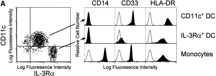
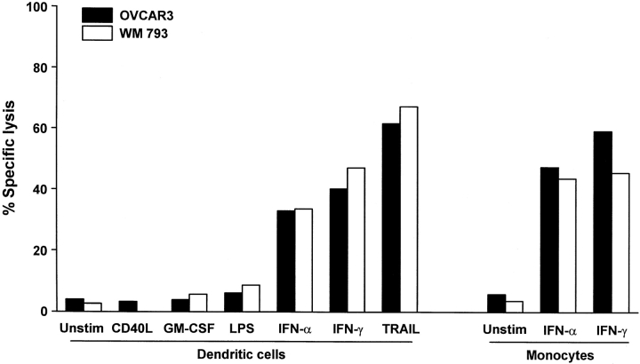
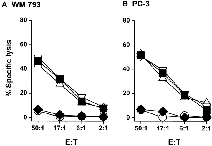
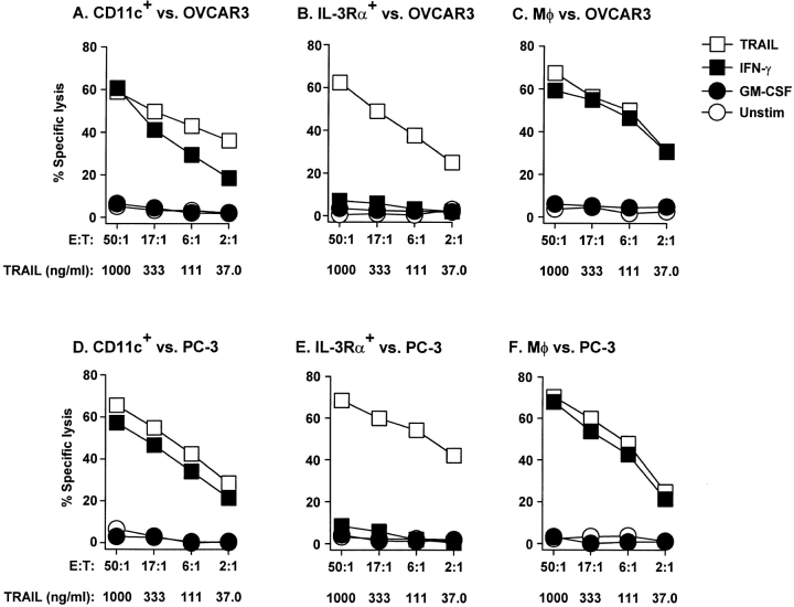
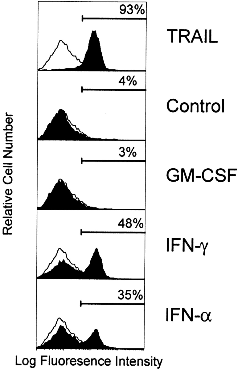
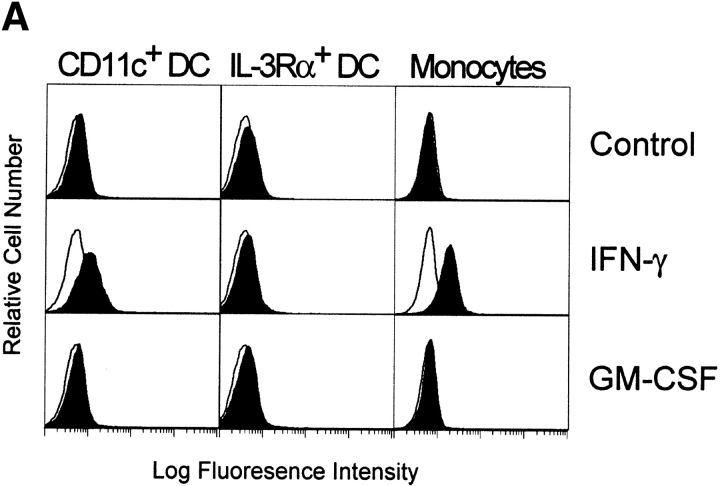
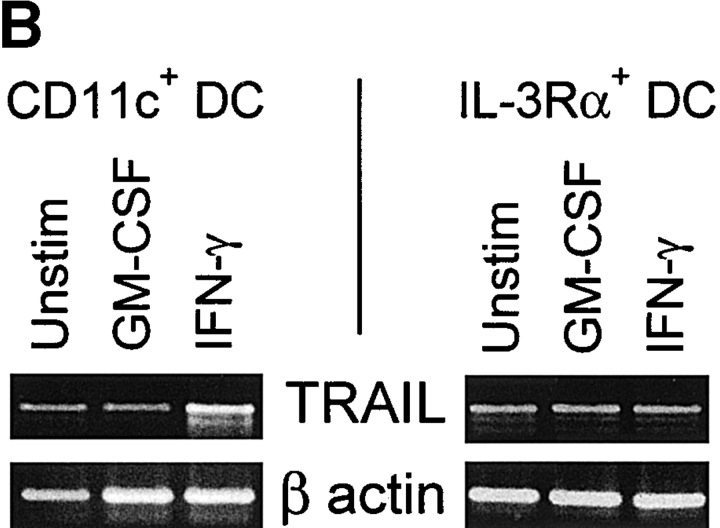
TRAIL (TNF-related apoptosis-inducing ligand) is a member of the TNF family that induces apoptosis in a variety of cancer cells. In this study, we demonstrate that human CD11c(+) blood dendritic cells (DCs) express TRAIL after stimulation with either interferon (IFN)-gamma or -alpha and acquire the ability to kill TRAIL-sensitive tumor cell targets but not TRAIL-resistant tumor cells or normal cell types. The DC-mediated apoptosis was TRAIL specific, as soluble TRAIL receptor blocked target cell death. Moreover, IFN-stimulated interleukin (IL)-3 receptor (R)alpha(+) blood precursor (pre-)DCs displayed minimal cytotoxicity toward the same target cells, demonstrating a clear functional difference between the CD11c(+) DC and IL-3Ralpha(+) pre-DC subsets. These results indicate that TRAIL may serve as an innate effector molecule on CD11c(+) DCs for the elimination of spontaneously arising tumor cells and suggest a means by which TRAIL-expressing DCs may regulate or eliminate T cells responding to antigen presented by the DCs.


Figures

Figure 1
Figure 1
Surface phenotype of freshly isolated CD11c+ and IL3Rα+ pre-DC subsets. (A) The two blood DCs were distinguishable based on CD11c and IL-3Rα expression as determined by multicolor flow cytometry. The CD11c+ and IL-3Rα+ pre-DCs were compared with Mφ for CD14, CD33, and HLA-DR expression. Filled histograms represent staining with specific antibody; open histograms represent isotype-matched controls. (B) CD11c+ DCs, but not IL-3Rα+ pre-DCs, express CD83 after 12-h incubation in complete medium. Histograms and contour plots represent 104 gated DCs, and viability was >95% as assessed by propidium iodide exclusion.
Figure 1
Figure 1
Surface phenotype of freshly isolated CD11c+ and IL3Rα+ pre-DC subsets. (A) The two blood DCs were distinguishable based on CD11c and IL-3Rα expression as determined by multicolor flow cytometry. The CD11c+ and IL-3Rα+ pre-DCs were compared with Mφ for CD14, CD33, and HLA-DR expression. Filled histograms represent staining with specific antibody; open histograms represent isotype-matched controls. (B) CD11c+ DCs, but not IL-3Rα+ pre-DCs, express CD83 after 12-h incubation in complete medium. Histograms and contour plots represent 104 gated DCs, and viability was >95% as assessed by propidium iodide exclusion.
Figure 2
Figure 2
Blood CD11c+ DCs and IL-3Rα+ pre-DCs exhibit distinct morphological differences. Wright-Giemsa staining of freshly sorted CD11c+ DCs (A) and IL-3Rα+ pre-DCs (B). Photomicrographs were captured at a magnification of 40.
Figure 3
Figure 3
Cytolytic activity by human DCs after stimulation. Bulk DCs were incubated for 12 h in the absence (Unstim.) or presence of CD40L, GM-CSF, LPS, IFN-α, or IFN-γ and then cultured for 8 h with 51Cr-labeled OVCAR3 or WM 793 target cells at an E/T ratio of 50:1. As a positive control, soluble TRAIL was added to target cells at 1 μg/ml. For comparison, the cytolytic activity of unstimulated (Unstim.), IFN-α–, or IFN-γ–stimulated human monocytes at 50:1 E/T is also included. Data represent the mean of triplicate wells, and experiments were repeated at least three times with similar results. SD bars were omitted from the graphs but were <10% of the value of all points.
Figure 5
Figure 5
Specificity of CD11c+ DC cytolytic activity is restricted to TRAIL. Inclusion of the fusion protein TRAILR2–Fc (20 μg/ml), but not Fas–Fc (20 μg/ml) or TNFR–Fc (20 μg/ml), to 12-h IFN-γ–stimulated CD11c+ DCs inhibited killing of WM 793 (A) or PC-3 (B) tumor cell targets. Data points represent the mean of triplicate wells, and experiments were repeated at least three times with similar results. For clarity, SD bars were omitted from the graphs but were <10% of the value of all points. ○, unstimulated; ▪, IFN-γ; ♦, IFN-γ + TR2–Fc; ▵, IFN-γ + Fas–Fc; ▿, IFN-γ + TNFR–Fc.
Figure 4
Figure 4
TRAIL-mediated tumoricidal activity of blood DCs is restricted to CD11c+ DCs. CD11c+ DCs, IL-3Rα+ pre-DCs, and Mφ were incubated for 12 h in the absence or presence of GM-CSF or IFN-γ and then cultured for 8 h with 51Cr-labeled OVCAR3 (A–C) or PC-3 (D–F) target cells at the indicated E/T ratios. As a positive control, soluble TRAIL was added to the target cells at the indicated concentrations. Data represent the mean of triplicate wells, and experiments were repeated at least three times with similar results. SD bars were omitted from the graphs but were <10% of the value of all points.
Figure 6
Figure 6
Tumor cell targets undergo apoptotic cell death when cultured with IFN-stimulated DCs as determined by phosphatidylserine externalization. OVCAR3 tumor cells were cultured for 8 h in complete medium alone or in the presence of LZ-TRAIL (1 μg/ml), unstimulated DCs, or cytokine (GM-CSF, IFN-γ, or IFN-α [100 ng/ml for 12 h])-stimulated DCs (E/T ratio 4:1). Cells were then stained with FITC–annexin V and analyzed by flow cytometry. The percentage of FITC–annexin V–positive tumor cells is indicated for each condition. Histograms represent 104 gated tumor cells. Similar results were seen with DCs from three other donors.
Figure 7
Figure 7
TRAIL expression on IFN-stimulated human CD11c+ DCs and Mφ but not IL-3Rα+ pre-DCs. (A) Flow cytometric analysis of TRAIL protein expression. DCs and Mφ were incubated for 12 h in the absence or presence of IFN-γ or GM-CSF and then analyzed for TRAIL expression. Open histograms represent staining by the FITC-labeled M181 (anti-TRAIL mAb); filled histograms represent staining by the FITC-labeled isotype control. Histograms represent 104 gated cells in all conditions. (B) RT-PCR analysis of TRAIL mRNA levels in CD11c+ and IL-3Rα+ pre-DCs. Sorted CD11c+ and IL-3Rα+ pre-DCs were incubated for 12 h in the absence or presence of IFN-γ or GM-CSF. β-actin was used as a control over the same time period.
Figure 7
Figure 7
TRAIL expression on IFN-stimulated human CD11c+ DCs and Mφ but not IL-3Rα+ pre-DCs. (A) Flow cytometric analysis of TRAIL protein expression. DCs and Mφ were incubated for 12 h in the absence or presence of IFN-γ or GM-CSF and then analyzed for TRAIL expression. Open histograms represent staining by the FITC-labeled M181 (anti-TRAIL mAb); filled histograms represent staining by the FITC-labeled isotype control. Histograms represent 104 gated cells in all conditions. (B) RT-PCR analysis of TRAIL mRNA levels in CD11c+ and IL-3Rα+ pre-DCs. Sorted CD11c+ and IL-3Rα+ pre-DCs were incubated for 12 h in the absence or presence of IFN-γ or GM-CSF. β-actin was used as a control over the same time period.

References

    1. Steinman R.M. The dendritic cell system and its role in immunogenicity. Annu. Rev. Immunol. 1991;9:271–296. - PubMed
    1. Banchereau J., Steinman R.M. Dendritic cells and the control of immunity. Nature. 1998;392:245–252. - PubMed
    1. O'Doherty U., Steinman R.M., Peng M., Cameron P.U., Gezelter S., Kopeloff I., Swiggard W.J., Pope M., Bhardwaj N. Dendritic cells freshly isolated from human blood express CD4 and mature into typical immunostimulatory dendritic cells after culture in monocyte-conditioned medium. J. Exp. Med. 1993;178:1067–1076. - PMC - PubMed
    1. O'Doherty U., Peng M., Gezelter S., Swiggard W.J., Betjes M., Bhardwaj N., Steinman R.M. Human blood contains two subsets of dendritic cells, one immunologically mature and the other immature. Immunology. 1994;82:487–493. - PMC - PubMed
    1. Nijman H.W., Kleijmeer M.J., Ossevoort M.A., Oorschot V.M.J., Vierboom M.P.M., van de Keur M., Kenemans P., Kast W.M., Geuze H.J., Melief C.J.M. Antigen capture and major histocompatibility class II compartments of freshly isolated and cultured human blood dendritic cells. J. Exp. Med. 1995;182:163–174. - PMC - PubMed

MeSH terms