. 1999 Oct 18;190(8):1165-74.
doi: 10.1084/jem.190.8.1165.

Protective T cell-independent antiviral antibody responses are dependent on complement

Affiliations

Protective T cell-independent antiviral antibody responses are dependent on complement

A F Ochsenbein et al. J Exp Med. .

Abstract

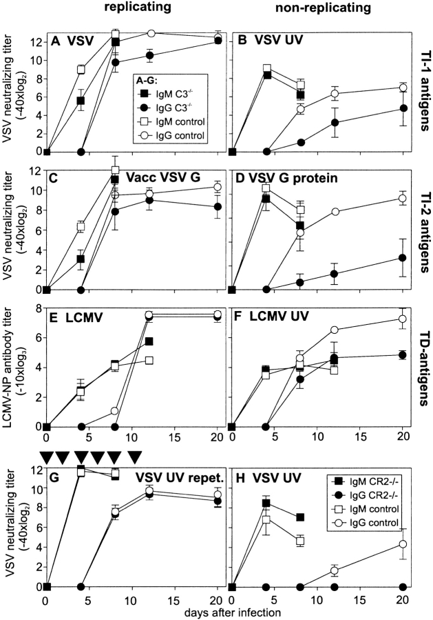
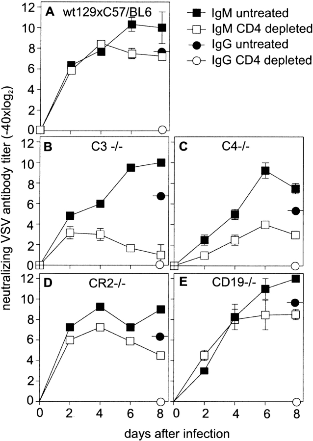
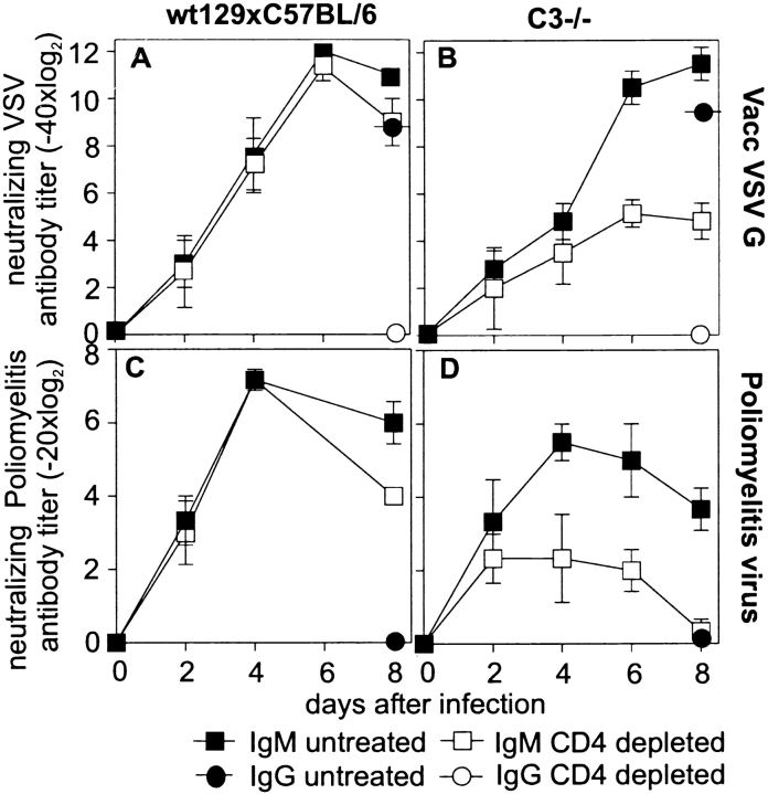
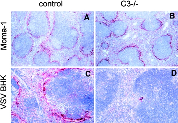
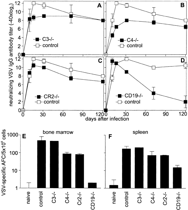
Complement is part of the innate immune system and one of the first lines of host defense against infections. Its importance was evaluated in this study in virus infections in mice deficient either in soluble complement factors (C3(-/-), C4(-/-)) or in the complement signaling complex (complement receptor [CR]2(-/-), CD19(-/-)). The induction of the initial T cell-independent neutralizing immunoglobulin (Ig)M antibody response to vesicular stomatitis virus (VSV), poliomyelitis virus, and recombinant vaccinia virus depended on efficient antigen trapping by CR3 and -4-expressing macrophages of the splenic marginal zone. Neutralizing IgM and IgG antibody responses were largely independent of CR2-mediated stimulation of B cells when mice were infected with live virus. In contrast, immunizations with nonreplicating antigens revealed an important role of B cell stimulation via CR2 in the switch to IgG. The complement cascade was activated after infection with VSV via the classical pathway, and active complement cleavage products augmented the effector function of neutralizing IgM and IgG antibodies to VSV by a factor of 10-100. Absence of the early neutralizing antibody responses, together with the reduced efficiency of neutralizing IgM in C3(-/-) mice, led to a drastically enhanced susceptibility to disease after infection with VSV.


Figures

Figure 1
Figure 1
Antibody responses to replicating and nonreplicating TI-1, TI-2, and TD viral antigens. C3−/− (▪, •) and control mice (□, ○) ([C57BL/6 × 129Sv]F1 or C57BL/6) were immunized with (A) 2 × 10⁶ pfu live VSV i.v., (B) 2 × 10⁸ pfu VSV UV-inactivated i.v., (C) 2 × 10⁶ pfu Vacc VSV G, or (D) 10 μg baculovirus VSV G protein i.v. Neutralizing antibody titers were assessed at the time points indicated. In the experiment shown, three out of five C3−/− mice died between day 8 and 12 after infection with VSV (Table ). Antibody titers in the dying mice were not different from those in surviving mice. After immunization with (E) 200 pfu LCMV-WE or (F) UV-inactivated purified LCMV-WE (∼20 μg protein), LCMV NP–specific antibodies were determined in an ELISA on baculovirus-derived LCMV NP–coated plates. Titers are shown as dilutions leading to OD 405 nm twice over background. (G) C3−/− and control mice were immunized every other day until day 12 with 2 × 10⁸ pfu UV-inactivated VSV, and neutralizing antibody titers were measured. (H) CR2−/− and control mice were immunized once with 2 × 10⁸ pfu UV-inactivated VSV, and VSV-IND–neutralizing antibody titers were assessed at the time points indicated. All results (A–H) are given as mean ± SD of three mice per group. Experiments were repeated twice with similar results.
Figure 2
Figure 2
T cell dependence of VSV-specific IgM antibody responses. Control (C57BL/6 × 129Sv)F1 (A), C3−/− (B), C4−/− (C), CR2−/− (D), and CD19−/− (E) were infected with 2 × 10⁶ pfu VSV. Total VSV-neutralizing Ig was assessed at the time points indicated. Mice were either depleted of CD4+ T cells (two 200-μl administrations of anti-CD4 antibody YTS191.1, on day 3 and day 1) or left untreated. Efficiency of CD4+ T cell depletion was checked by FACS™. Neutralizing IgG (circles) was assessed on day 8 (reduction with β-ME) and was always at least two titer steps lower than total Ig, indicating that antibody titers up to day 8 represent IgM. One of three comparable experiments is shown.
Figure 3
Figure 3
TD IgM antibody responses to recombinant vaccinia virus and poliomyelitis virus. C3−/− and control mice (either depleted of CD4+ T cells or left untreated) were immunized with 2 × 10⁶ pfu Vacc VSV G (A and B), and neutralizing antibody titers were assessed every other day. Neutralizing IgG (circles) was determined 8 d after immunization and was always at least two titer steps lower than total Ig, indicating that antibody titers until day 8 represent IgM. CD4+ T cell–depleted and untreated control (C) or C3−/− (D) mice were immunized with 500 μl of inactivated polio vaccine (Salk), and polio virus (serotype II)-specific neutralizing antibodies were assessed until day 8. No switch to IgG was observed after single immunization with polio vaccine. The experiment was repeated twice with comparable results.
Figure 4
Figure 4
Recruitment of viral antigen to the spleen. The spleens of naive (C57BL/6 × 129Sv)F1 (A, control) and C3−/− (B) mice were stained with MOMA-1 (specific for metallophilic macrophages of the marginal zone). Control and C3−/− mice were immunized intraperitoneally with viral antigen derived from disrupted VSV-infected BHK cells. 1 d later, VSV antigen was stained on spleen sections as described in Materials and Methods (original magnification: A and B, 125; C and D, 250). One of two comparable experiments is shown.
Figure 5
Figure 5
Long-term antibody and B cell memory in complement-deficient mice. C3−/− (A), C4−/− (B), CR2−/− (C), and CD19−/− (D) mice were immunized with 2 × 10⁶ pfu VSV, and long-term antibody titers were compared with controls. Three of six C3−/− and two of five C4−/− animals died between day 8 and 12 after immunization. Antibody titers in surviving and dying mice were comparable until day 8. Antibody titers in surviving animals were followed up to day 120. 120 d after infection, VSV-specific AFCs in the spleen (F) and bone marrow (E) were assessed in an enzyme-linked immunospot assay. Results are given as mean ± SD of three mice per group. Experiments were repeated twice with comparable results.

References

    1. Wessels M.R., Butko P., Ma M., Warren H.B., Lage A.L., Carroll M.C. Studies of group B streptococcal infection in mice deficient in complement component C3 or C4 demonstrate an essential role for complement in both innate and acquired immunity. Proc. Natl. Acad. Sci. USA. 1995;92:11490–11494. - PMC - PubMed
    1. Holland S.M., Gallin J.I. Evaluation of the patient with recurrent bacterial infections. Annu. Rev. Med. 1998;49:185–199. - PubMed
    1. Lachmann P.J., Davies A. Complement and immunity to viruses. Immunol. Rev. 1997;159:69–77. - PubMed
    1. Stoiber H., Clivio A., Dierich M.P. Role of complement in HIV infection. Annu. Rev. Immunol. 1997;15:649–674. - PubMed
    1. Carter R.H., Fearon D.T. CD19lowering the threshold for antigen receptor stimulation of B lymphocytes. Science. 1992;256:105–107. - PubMed

Publication types

MeSH terms