Comparative Study
. 1999 Nov;19(11):7336-46.
doi: 10.1128/MCB.19.11.7336.

Eukaryotic translation initiation factor 4AIII (eIF4AIII) is functionally distinct from eIF4AI and eIF4AII

Affiliations
Comparative Study

Eukaryotic translation initiation factor 4AIII (eIF4AIII) is functionally distinct from eIF4AI and eIF4AII

Q Li et al. Mol Cell Biol. 1999 Nov.

Abstract

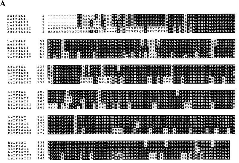
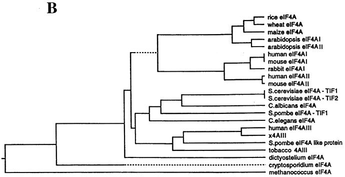
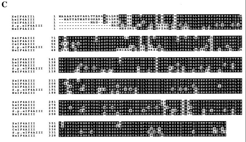
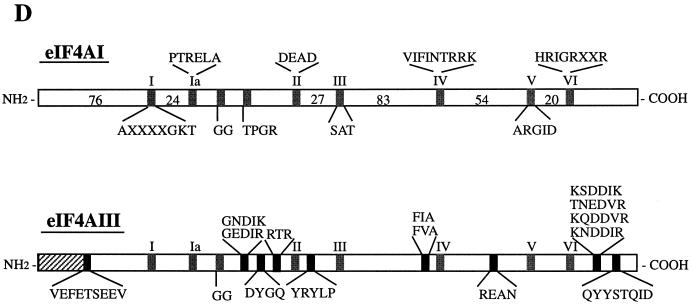
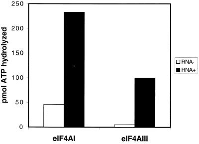
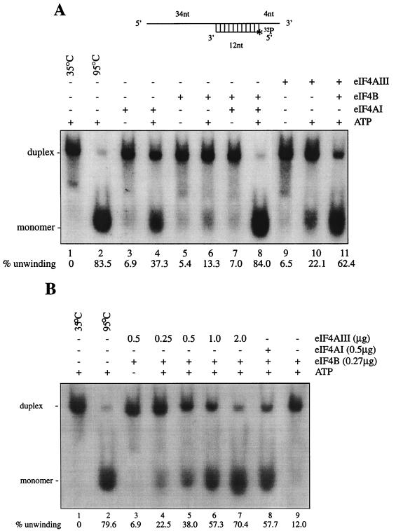
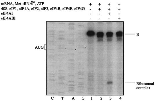
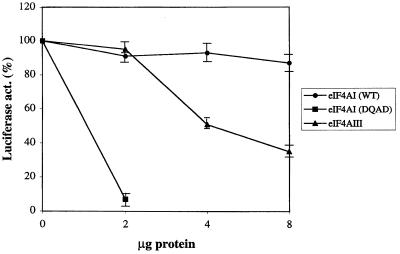
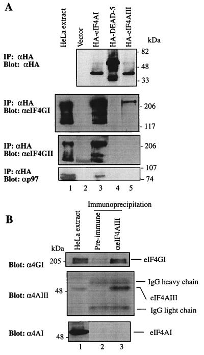
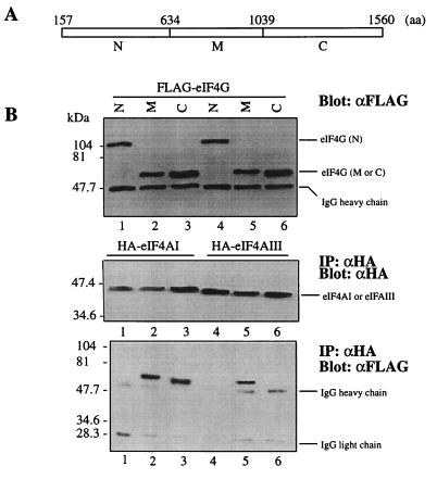
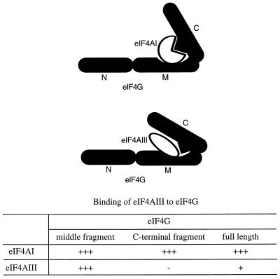
Eukaryotic initiation factor 4A (eIF4A) is an RNA-dependent ATPase and ATP-dependent RNA helicase that is thought to melt the 5' proximal secondary structure of eukaryotic mRNAs to facilitate attachment of the 40S ribosomal subunit. eIF4A functions in a complex termed eIF4F with two other initiation factors (eIF4E and eIF4G). Two isoforms of eIF4A, eIF4AI and eIF4AII, which are encoded by two different genes, are functionally indistinguishable. A third member of the eIF4A family, eIF4AIII, whose human homolog exhibits 65% amino acid identity to human eIF4AI, has also been cloned from Xenopus and tobacco, but its function in translation has not been characterized. In this study, human eIF4AIII was characterized biochemically. While eIF4AIII, like eIF4AI, exhibits RNA-dependent ATPase activity and ATP-dependent RNA helicase activity, it fails to substitute for eIF4AI in an in vitro-reconstituted 40S ribosome binding assay. Instead, eIF4AIII inhibits translation in a reticulocyte lysate system. In addition, whereas eIF4AI binds independently to the middle and carboxy-terminal fragments of eIF4G, eIF4AIII binds to the middle fragment only. These functional differences between eIF4AI and eIF4AIII suggest that eIF4AIII might play an inhibitory role in translation under physiological conditions.


Figures

FIG. 1
FIG. 1
(A) Protein sequence alignment of eIF4A proteins. The pattern-induced multisequence alignment program was used to align the amino acid sequences of human (h), mouse (m), and Xenopus (X) eIF4A proteins. Identical and conserved amino acid residues are in black and shaded boxes, respectively. (B) Phylogenetic relationship among members of the eIF4A gene family based on aligned amino acid sequences, which was created by the PILEUP program. (C) Sequence alignment of human (h), Xenopus (X), S. pombe (S.p.), C. elegans (C), and Nicotiana plumbaginifolia (N) eIF4AIII. The accession numbers are as follows: heIF4AI (P04765), meIF4AI (S00986), heIF4AII (D30655), meIF4AII (S00985), XeIF4AIII (AAB71410), S.P.eIF4AIII (CAA92238), CeIF4AIII (AAB96704), and NeIF4AIII (P41380). (D) Amino acid motifs which are conserved in the eIF4A family and which are specific for eIF4AIII. The N-terminal divergent sequence in eIF4AIII is hatched.
FIG. 1
FIG. 1
(A) Protein sequence alignment of eIF4A proteins. The pattern-induced multisequence alignment program was used to align the amino acid sequences of human (h), mouse (m), and Xenopus (X) eIF4A proteins. Identical and conserved amino acid residues are in black and shaded boxes, respectively. (B) Phylogenetic relationship among members of the eIF4A gene family based on aligned amino acid sequences, which was created by the PILEUP program. (C) Sequence alignment of human (h), Xenopus (X), S. pombe (S.p.), C. elegans (C), and Nicotiana plumbaginifolia (N) eIF4AIII. The accession numbers are as follows: heIF4AI (P04765), meIF4AI (S00986), heIF4AII (D30655), meIF4AII (S00985), XeIF4AIII (AAB71410), S.P.eIF4AIII (CAA92238), CeIF4AIII (AAB96704), and NeIF4AIII (P41380). (D) Amino acid motifs which are conserved in the eIF4A family and which are specific for eIF4AIII. The N-terminal divergent sequence in eIF4AIII is hatched.
FIG. 2
FIG. 2
Northern blot analysis of human eIF4AIII mRNA expression in human tissues. A multiple-tissue Northern blot, MTN1 (A) and MTN2 (B) (CLONTECH), containing 2 μg of poly(A)+ RNA per lane from the indicated human tissues was hybridized with a 32P-labeled human eIF4AIII (Nuk34) cDNA probe (a 0.3-kb fragment from the 3′ untranslated sequence), as specified by the manufacturer. The filters were also hybridized with a [32P]-labeled actin probe. Taken from reference .
FIG. 3
FIG. 3
Levels of eIF4AIII protein. (A) Coomassie blue staining. Recombinant eIF4AI and eIF4AIII (1 μg) were resolved by SDS-PAGE (10% polyacrylamide). Molecular masses (in kilodaltons) of protein standards are indicated. (B) Immunological identification of eIF4AI and eIF4AIII. Recombinant eIF4AI (1 μg; lanes 1 and 3) and eIF4AIII (1 μg; lanes 2 and 4) were subjected to SDS-PAGE (10% polyacrylamide), and proteins were transferred onto a nitrocellulose membrane, which was probed with anti-eIF4AI (lanes 1 and 2) or anti-eIF4AIII (lanes 3 and 4) antibodies. Protein bands were visualized on an X-ray film by an enhanced chemiluminescence detection system. (C) Cell extracts (10 μg) were resolved by SDS-PAGE (10% polyacrylamide). Western blotting was performed with anti-eIF4AI or anti-eIF4AIII. The same membrane was reprobed with anti-actin antibody (bottom).
FIG. 4
FIG. 4
Quantitation of endogenous eIF4AIII and eIF4AI in HeLa cells. The indicated amounts of cell extract and amounts of eIF4AIII (A) or eIF4AI (B) were mixed and resolved by SDS-PAGE (10% polyacrylamide). Proteins were electroblotted onto a nitrocellulose membrane and probed with a polyclonal anti-eIF4AIII (A) or monoclonal eIF4AI (B) antibody. Signals were quantified on a phosphorimager, and the values obtained are indicated at the bottom of the figure.
FIG. 5
FIG. 5
eIF4AIII possesses an RNA-stimulated ATPase activity. ATPase assays were performed for 15 min with 100 μM ATP and 1 μg of recombinant eIF4AIII or recombinant eIF4AI in the absence or presence of 0.3 absorbance at 260 nm units of poly(A) RNA. The amount of inorganic phosphate released from a reaction with no eIF4A and no poly(A) RNA was subtracted. The values are the mean of three independent experiments.
FIG. 6
FIG. 6
RNA helicase activity of eIF4AIII. (A) An RNA duplex was used for the helicase assay, as described in Materials and Methods. eIF4AI (0.38 μg) or eIF4AIII (0.38 μg) was incubated in the presence or absence of eIF4B (0.23 μg) with 20,000 cpm (0.04 pmol) of 32P-labeled duplex RNA for 15 min at 35°C. (B) Dose-dependent helicase activity of eIF4AIII in the presence of eIF4B. nt, nucleotides.
FIG. 7
FIG. 7
eIF4AIII does not substitute for eIF4AI in the ribosome binding assay. The indicated translation components were incubated with β-globin mRNA. Formation of the 48S ribosomal complex was detected by toeprinting. Full-length cDNA is marked E. The cDNA product that maps to 15 to 17 nucleotides downstream of the initiation codon of β-globin mRNA is labeled Ribosomal complex. The position of the initiation codon is shown to the left of the reference lanes, which represent the β-globin sequence obtained with the same primer as for the toeprinting.
FIG. 8
FIG. 8
eIF4AIII inhibits translation. Increasing amounts of recombinant eIF4AI (2 to 8 μg), eIF4AI mutant (DQAD) (2 μg) (28), or eIF4AIII (2 to 8 μg) were preincubated in a rabbit reticulocyte lysate for 5 min at 30°C. Uncapped uciferase mRNA was added to the lysate, and translation was carried out at 37°C for 30 min. The luciferase activity of the lysate preincubated with buffer alone was set at 100%. Each point represents the mean of four experiments. WT, wild type.
FIG. 9
FIG. 9
eIF4AIII interacts with eIF4G. (A). Expression plasmids pcDNA3-HA, pcDNA3-HA-eIF4AI, pcDNA3-HA-DEAD5, and pcDNA3-HA-eIF4AIII were transfected into HeLa cells after infection with vTF7-3, as described in Materials and Methods. Immunoprecipitates (IP) obtained with anti-HA antibody (αHA) were resolved by SDS-PAGE (10% polyacrylamide). Western blotting was performed with anti-HA (top), anti-eIF4GI (αeIF4GI) (second panel), anti-eIF4GII (αeIF4GII) (third panel), or anti-p97 antibody (αp97) (bottom). (B) HeLa cell extract (168 μg) was incubated with preimmune serum or anti-eIF4AIII serum. Following precipitation with protein G-Sepharose, bound proteins were resolved by SDS-PAGE (10% polyacrylamide). Western blotting was performed with anti-eIF4GI (top), anti-eIF4AIII (middle), or anti-eIF4AI antibody (bottom). HeLa cell extract (12 μg) was used for Western blotting (lane 1). IgG, immunoglobulin G.
FIG. 10
FIG. 10
eIF4AIII binds only to the middle portion of eIF4GI. (A) Schematic map of the three fragments of eIF4GI. (B) FLAG-eIF4GI N-terminal region (N) (lanes 1 and 4), middle region (M) (lanes 2 and 5), and C-terminal region (C) (lanes 3 and 6) expression plasmids were cotransfected into HeLa cells with pcDNA3-HA-eIF4AI (lanes 1 to 3) or pcDNA3-HA-eIF4AIII (lanes 4 to 6). An aliquot of the extract was removed for Western blotting with anti-FLAG (top). The remaining portion was used for immunoprecipitation (IP) with anti-HA antibody (αHA). Western blotting of immunoprecipitates was performed with anti-HA (αHA) (middle) or anti-FLAG (αFLAG) (bottom) antibody.
FIG. 11
FIG. 11
Model of binding of eIF4AI or eIF4AIII to eIF4GI. While eIF4AI binds to the middle and C-terminal regions of eIF4GI, eIF4AIII binds only to the middle region. It should be noted that there is a distinct possibility that two molecules of eIF4AI bind to one molecule of eIF4G. N, N-terminal fragment; M, middle fragment; C, C-terminal fragment.

Similar articles

Cited by

References

    1. Allen M L, Metz A M, Timmer A T, Rhoads R E, Browning K S. Isolation and sequence of the cDNAs encoding the subunits of the isozyme form of wheat protein synthesis initiation factor 4F. J Biol Chem. 1992;267:23232–23236. - PubMed
    1. Audet R G, Goodchild J, Richter J D. Eukaryotic initiation factor 4A stimulates translation in microinjected Xenopus oocyte. Dev Biol. 1987;121:58–68. - PubMed
    1. Belsham G J, Sonenberg N. RNA-protein interactions in regulation of picornavirus RNA translation. Microbiol Rev. 1996;60:499–511. - PMC - PubMed
    1. Beretta L, Gingras A C, Svitkin Y V, Hall M N, Sonenberg N. Rapamycin blocks the phosphorylation of 4E-BP1 and inhibits cap-dependent initiation of translation. EMBO J. 1996;15:658–664. - PMC - PubMed
    1. Blum S, Schmid S R, Pause A, Buser P, Linder P, Sonenberg N, Trachsel H. ATP hydrolysis by initiation factor 4A is required for translation initiation in Saccharomyces cerevisiae. Proc Natl Acad Sci USA. 1992;89:7664–7668. - PMC - PubMed

Publication types

MeSH terms

Associated data

LinkOut - more resources