. 1999 Nov;19(11):7577-88.
doi: 10.1128/MCB.19.11.7577.

PBX and MEIS as non-DNA-binding partners in trimeric complexes with HOX proteins

Affiliations

PBX and MEIS as non-DNA-binding partners in trimeric complexes with HOX proteins

K Shanmugam et al. Mol Cell Biol. 1999 Nov.

Abstract

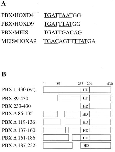
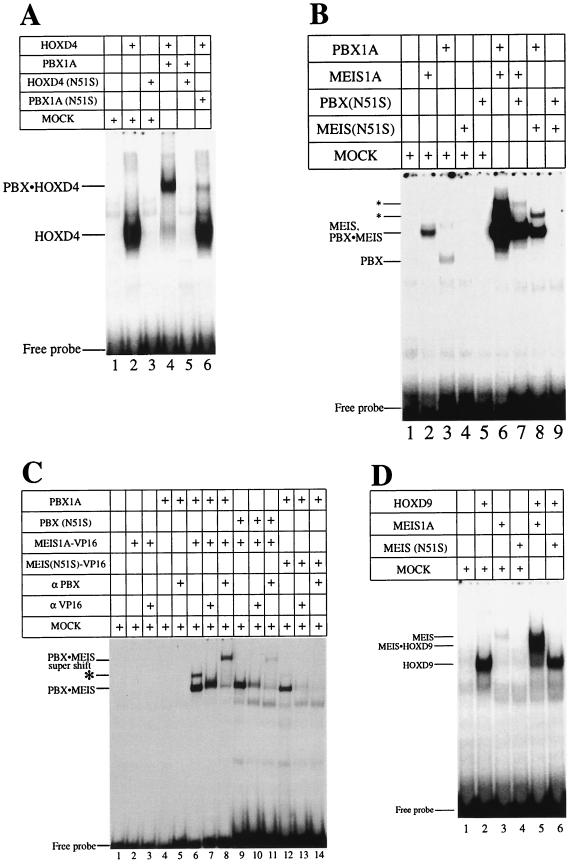
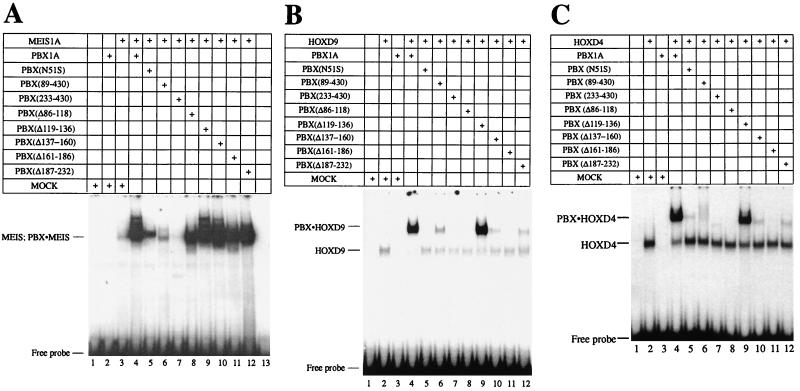
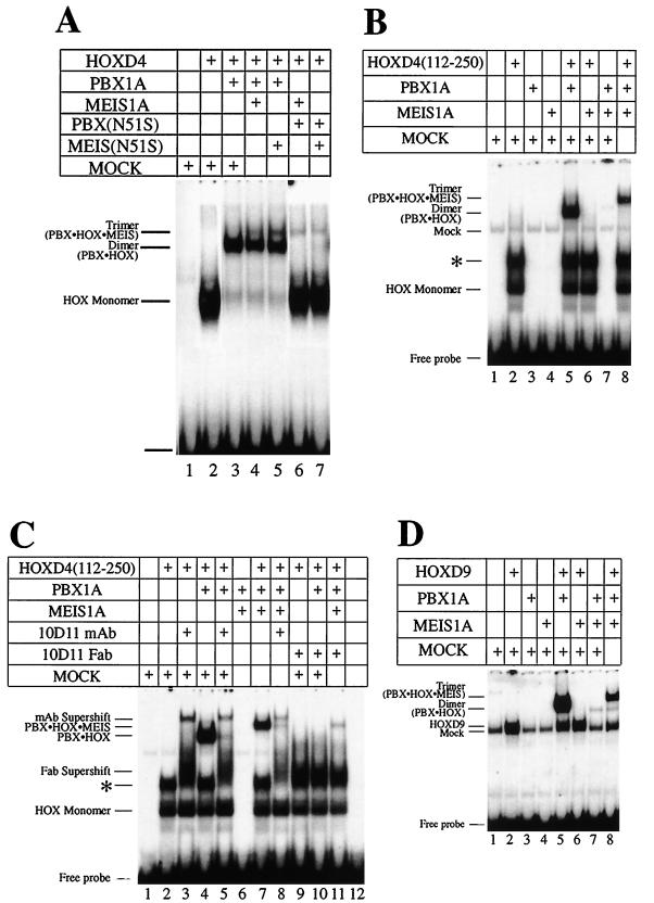
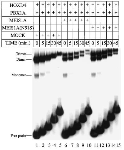
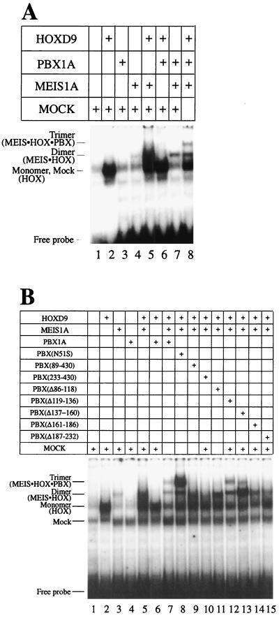
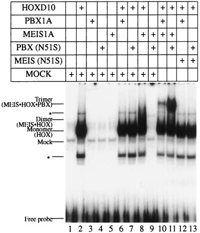
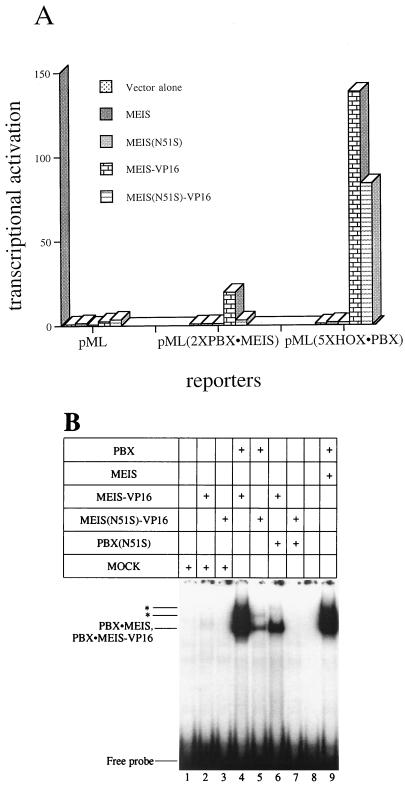
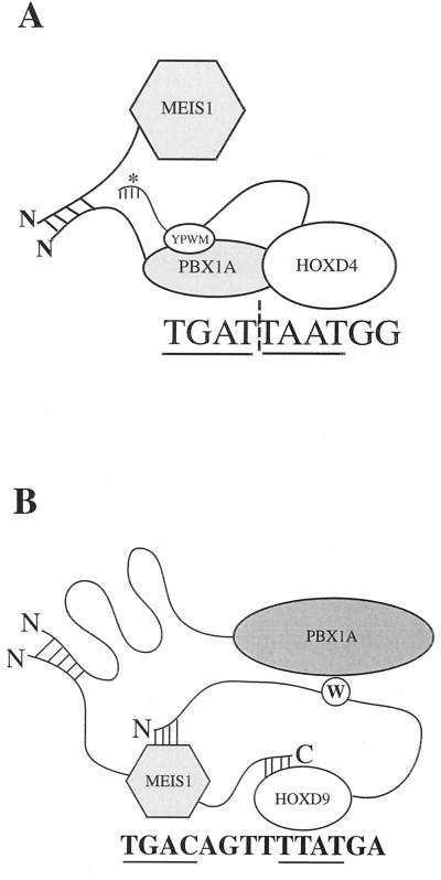
HOX, PBX, and MEIS transcription factors bind DNA through a homeodomain. PBX proteins bind DNA cooperatively as heterodimers with MEIS family members and also with HOX proteins from paralog groups 1 to 10. MEIS proteins cooperatively bind DNA with ABD-B class HOX proteins of groups 9 and 10. Here, we examine aspects of dimeric and higher-order interactions between these three homeodomain classes. The most significant results can be summarized as follows. (i) Most of PBX N terminal to the homeodomain is required for efficient cooperative binding with HOXD4 and HOXD9. (ii) MEIS and PBX proteins form higher-order complexes on a heterodimeric binding site. (iii) Although MEIS does not cooperatively bind DNA with ANTP class HOX proteins, it does form a trimer as a non-DNA-binding partner with DNA-bound PBX-HOXD4. (iv) The N terminus of HOXD4 negatively regulates trimer formation. (v) MEIS forms a similar trimer with DNA-bound PBX-HOXD9. (vi) A related trimer (where MEIS is a non-DNA-binding partner) is formed on a transcriptional promoter within the cell. (vii) We observe an additional trimer class involving non-DNA-bound PBX and DNA-bound MEIS-HOXD9 or MEIS-HOXD10 heterodimers that is enhanced by mutation of the PBX homeodomain. (viii) In this latter trimer, PBX is likely to contact both MEIS and HOXD9/D10. (ix) The stability of DNA binding by all trimers is enhanced relative to the heterodimers. These findings suggest novel functions for PBX and MEIS in modulating the function of DNA-bound MEIS-HOX and PBX-HOX heterodimers, respectively.


Figures

FIG. 1
FIG. 1
(A) Nucleotide sequences of the target sites used in this study. Position 6 is bold in both the PBX-HOXD4 and PBX-HOXD9 target sites. Core recognition elements for homeodomain binding are underlined. (B) Schematic representation of wild-type (wt) PBX1A and the different N-terminal and internal deletions used in this study. The homeodomain (HD) in PBX1A is comprised of aa 233 to 295.
FIG. 2
FIG. 2
DNA-binding activity of both partners contributes to cooperative heterodimer formation in EMSA. Wild-type or N51S mutants of HOXD4, PBX1A, and MEIS1A were tested for the ability to form cooperative heterodimers. (A) PBX-HOXD4 interaction. Full-length HOXD4 bound as a monomer (lane 2) and a cooperative heterodimer with PBX1A (lane 4) on a PBX-HOX consensus site (Fig. 1A). Mutating asparagine 51 in the homeodomain of HOXD4 resulted in the loss of monomeric DNA binding and heterodimeric DNA binding with PBX1A (lanes 3 and 5). Lane 6 shows that a similar mutation in the homeodomain of PBX resulted in the formation of a reduced cooperative complex. (B) PBX-MEIS interaction. MEIS and PBX bound weakly as monomers (lanes 2 and 3) to a PBX-MEIS site (Fig. 1A) but together formed a strong cooperative complex (lane 6). Mutating asparagine 51 in the homeodomain of MEIS or PBX resulted in the loss of monomer binding activity (lanes 4 and 5) and a significant reduction in the formation of the DNA-bound heterodimeric complex (lanes 7 and 8). Incubating MEIS(N51S) with PBX(N51S) results in the complete loss of the cooperative complex (lane 9). Asterisks denote two higher-order complexes of PBX and MEIS. (C) Dimeric and higher-order complexes that form on a PBX-MEIS binding site (lane 6) contain both PBX1A and a MEIS-VP16 fusion protein, as shown by the use of N51S mutants (lanes 9 and 12) and antibodies to PBX1A (lanes 8, 11, and 14) and VP16 (lanes 7, 10, and 13). Note that lanes 9 to 14 were exposed longer than lanes 1 to 8. The asterisk denotes the faster of the two higher-order complexes also seen in panel B. The slowest species is also visible with a longer exposure. (D) DNA binding by MEIS is indispensable for the formation of a MEIS-HOXD9 cooperative complex. HOXD9 bound well as a monomer and a cooperative dimer with MEIS1A to a MEIS-HOX site (lanes 2 and 5). Poor MEIS binding (lane 3) was lost upon mutating asparagine 51 to serine (lane 4). Incubating MEIS(N51S) in the presence of HOXD9 also resulted in the loss of the cooperative heterodimer. Mock lysate was used to normalize the levels of lysate in the reactions where one of the translated proteins was missing.
FIG. 3
FIG. 3
Mapping domains of PBX important for cooperative interactions. (A) Interaction of PBX1A with MEIS1A. Deletions encompassing region 1–232 of PBX1A (Fig. 1B) were assayed for the ability to interact with MEIS1A on a PBX-MEIS site in EMSA. The majority of the PBX1A-MEIS1A complex was abolished in the presence of PBX(89–430) (lane 6). Deleting the entire region N terminal to the PBX homeodomain [PBX(233-430)] resulted in the complete loss of the cooperative complex (lane 7). A series of deletions covering aa 86 to 232 did not reveal changes in the formation of the cooperative complex (lanes 8 to 12). Lane 1 served as a mock control. (B) Interaction of PBX1A with HOXD9. EMSA was performed as for panel A except that HOXD9 was used in the place of MEIS1A. PBX1A and deleted derivatives (Fig. 1B) were analyzed for the ability to form a cooperative complex with HOXD9 on a T6 probe (Fig. 1A). Except PBX(Δ119-136) (lane 9), all deletions of PBX had a profound impact on the formation of a cooperative complex (lanes 5 to 11). Lane 1 served as a mock control; lanes 2 and 4 show the DNA-bound HOXD9 monomer and PBX-HOXD9 cooperative complex, respectively. (C) As for panel B but with HOXD4 replacing HOXD9.
FIG. 4
FIG. 4
Formation of a trimeric complex by MEIS(N51S). (A) MEIS1A is the non-DNA-binding component in the trimer with PBX-HOXD4 as seen in EMSA. Full-length HOXD4 binds an A6 site (Fig. 1A) as a monomer (lane 2) and as a cooperative dimer with PBX (lane 3). Addition of MEIS1A or MEIS(N51S) leads to the formation of an additional low-mobility complex (lanes 4 and 5). Loss of the cooperative dimer and retention of the presumptive trimer were observed when PBX(N51S) was used in the presence of MEIS1A and HOXD4 or of MEIS(N51S) and HOXD4 (lanes 6 and 7). (B) Deletion of aa 1 to 111 from HOXD4 reveals a negative effect of this region on the formation of the trimer. Deleting the first 111 aa resulted in a more robust trimeric complex: all of the heterodimer is converted to presumptive heterotrimer in lane 8 (compare to lane 4 in Fig. 4A). Also compare lanes 4 and 7 in Fig. 1C. (C) A MAb against the HOXD4 YPWM prevents the formation of the trimeric complex in EMSA. Addition of MAb 10D11 (54) or the respective Fab fragments led to the loss of the dimeric and trimeric complexes (compare lane 5 to lane 4, lane 8 to lane 7, and lanes 10 and 11 to lanes 4 and 7). (D) MEIS1A can be a non-DNA-binding component in a trimer with PBX1A-HOXD9. Addition of MEIS1A to PBX1A and HOXD9 led to the formation of a low-mobility band in EMSA (compare lane 8 to lane 5). On the site used here, no or little DNA-binding activity was observed for MEIS1A as a monomer (lane 4) or in combination with HOXD9 (lane 6) or PBX1A (lane 7), suggesting that DNA binding by MEIS1A is not involved in the formation of the presumptive trimer. ∗, an apparent dimer of HOXD4 that we have shown to be very unstable (data not shown). A 32P-labeled T6 target site (Fig. 1A) was used as a probe.
FIG. 5
FIG. 5
MEIS1A and MEIS(N51S) can stabilize a trimeric complex as a non-DNA-binding partner. Dissociation rates were determined on an A6 labeled probe in the presence of a 100-fold excess of A6 cold competitor for the following complexes: PBX1A-HOXD4 (lanes 1 to 5), PBX1A-HOXD4 and MEIS1A-PBX1A-HOXD4 (lanes 6 to 10), and PBX1A-HOXD4 and MEIS(N51S)-PBX1A-HOXD4 (lanes 11 to 15).
FIG. 6
FIG. 6
PBX1A is the non-DNA-binding partner in a trimeric complex with MEIS1A and HOXD9. (A) PBX1A forms a trimeric complex with MEIS1A and HOXD9 in EMSA. Addition of PBX1A to MEIS1A and HOXD9 results in the decrease of monomer (lane 2) and dimer (lane 5) bands and the formation of a new low-mobility band (lane 8). (B) PBX1A is the non-DNA-binding partner in the trimeric complex and contacts both MEIS1A and HOXD9. This is revealed by the improved trimer formation with PBX(N51S) (lane 8) and reduced trimer formation with most N-terminal mutants of PBX1A (lanes 9 to 15 and Fig. 1B). A MEIS-HOX consensus binding site (Fig. 1A) (57) was used as probe in both panels.
FIG. 7
FIG. 7
PBX1A can form a trimeric complex with MEIS1A and HOXD10 as a non-DNA-binding partner in EMSA. PBX1A or PBX(N51S) forms a trimeric complex with MEIS1A and HOXD10 on a MEIS-HOX binding site (lanes 10 and 11). ∗, minor band resulting from the in vitro translation of HOXD10.
FIG. 8
FIG. 8
MEIS(N51S) can form a trimeric complex with HOX and PBX in vivo. (A) Effects of transfected expression vectors for MEIS1A, MEIS(N51S), MEIS-VP16, or MEIS(N51S)-VP16 on luciferase reporters containing no specific binding sites (pML) or binding sites for PBX-MEIS [pML(2×PBX · MEIS)] or HOX-PBX [pML(5×HOX · PBX)]. Results were reproducible, and data from one experiment are presented. Values are expressed as fold activation over transfection of the respective reporter plasmids alone. (B) Characterization of MEIS-VP16 and MEIS(N51S)-VP16 fusion proteins in EMSA. MEIS-VP16 or MEIS(N51S)-VP16 used for panel A were able to form complexes with PBX1A to the same degree as their wild-type counterparts (compare lanes 4, 5, and 9). ∗, higher-order PBX-MEIS complex also seen in Fig. 2B.
FIG. 9
FIG. 9
Model summarizing the formation of trimeric complexes by HOX cofactors MEIS and PBX as non-DNA-binding partners. (A) MEIS as the non-DNA-binding partner. PBX and HOX bind cooperatively to a consensus heterodimer binding site. MEIS and PBX interact through their N termini. ∗, deletion of the N terminus of HOXD4 results in a more robust trimeric complex. The significance of this effect is discussed in the text. (B) PBX as the non-DNA-binding partner. MEIS and HOXD9 or HOXD10 cooperatively bind a consensus MEIS-HOX binding site. We propose that PBX and MEIS interact via their N termini and that the PBX homeodomain contacts the tryptophan motif in HOXD9. The N terminus of PBX is looped to suggest the presence of intramolecular interactions important for the formation of the PBX-HOXD9 dimer and MEIS-HOXD9-PBX trimer. The importance of the N51S mutation in PBX and MEIS (not shown) is addressed in the Discussion. The core binding sites in both panels are underlined.

References

    1. Abu-Shaar M, Ryoo H D, Mann R S. Control of the nuclear localization of Extradenticle by competing nuclear import and export signals. Genes Dev. 1999;13:935–945. - PMC - PubMed
    1. Andrew S M, Titus J A. Fragmentation of immunoglobulin G. New York, N.Y: Greene Publishing Associates and Wiley Interscience; 1992.
    1. Bergson C, McGinnis W. An autoregulatory enhancer element of the Drosophila homeotic gene Deformed. EMBO J. 1990;9:4287–4297. - PMC - PubMed
    1. Berthelsen J, Kilstrup-Nielsen C, Blasi C, Mavilio F, Zappavigna V. The subcellular localization of PBX1 and EXD proteins depends on nuclear import and export signals and is modulated by association with PREP1 and HTH. Genes Dev. 1999;13:946–953. - PMC - PubMed
    1. Berthelsen J, Zappavigna V, Ferretti E, Mavilio F, Blasi F. The novel homeoprotein Prep1 modulates Pbx-Hox protein cooperativity. EMBO J. 1998;17:1434–1445. - PMC - PubMed

Publication types

MeSH terms

Substances

LinkOut - more resources