. 1999 Nov;19(11):7782-91.
doi: 10.1128/MCB.19.11.7782.

Evidence for distinct substrate specificities of importin alpha family members in nuclear protein import

Affiliations

Evidence for distinct substrate specificities of importin alpha family members in nuclear protein import

M Köhler et al. Mol Cell Biol. 1999 Nov.

Abstract

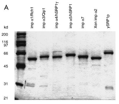
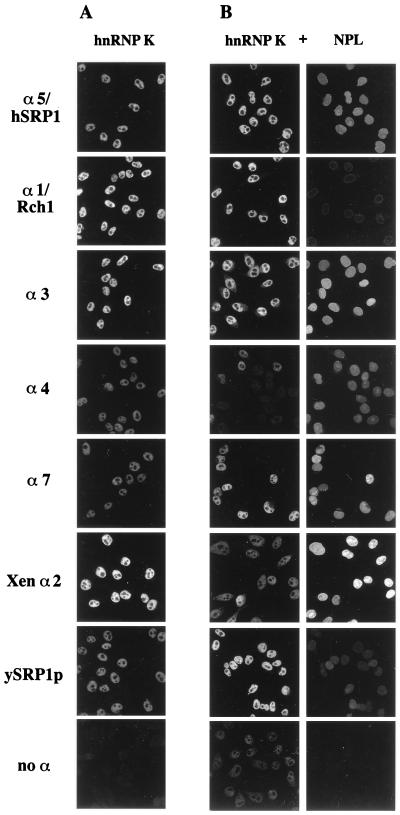
Importin alpha plays a pivotal role in the classical nuclear protein import pathway. Importin alpha shuttles between nucleus and cytoplasm, binds nuclear localization signal-bearing proteins, and functions as an adapter to access the importin beta-dependent import pathway. In contrast to what is found for importin beta, several isoforms of importin alpha, which can be grouped into three subfamilies, exist in higher eucaryotes. We describe here a novel member of the human family, importin alpha7. To analyze specific functions of the distinct importin alpha proteins, we recombinantly expressed and purified five human importin alpha's along with importin alpha from Xenopus and Saccharomyces cerevisiae. Binding affinity studies showed that all importin alpha proteins from humans or Xenopus bind their import receptor (importin beta) and their export receptor (CAS) with only marginal differences. Using an in vitro import assay based on permeabilized HeLa cells, we compared the import substrate specificities of the various importin alpha proteins. When the substrates were tested singly, only the import of RCC1 showed a strong preference for one family member, importin alpha3, whereas most of the other substrates were imported by all importin alpha proteins with similar efficiencies. However, strikingly different substrate preferences of the various importin alpha proteins were revealed when two substrates were offered simultaneously.


Figures

FIG. 1
FIG. 1
(A) Amino acid sequence of human importin α7. Residues in human importin α6 and mouse importin α-S2 that differ from those in human importin α7 are indicated above or below the importin α7 sequence, respectively. Epitopes used for antibody generation are underlined. (B) Alignment tree of all known importin α homologues. Tree construction was performed with the CLUSTAL program. All putative complete proteins of the GenBank and Swiss-Prot databases containing arm repeats and an IBB domain were included in the analysis. The accession numbers of the proteins are (Imp, importin): Imp α homologue (hom.), rice, AB006788; Imp α homologue A, C. eleg., gi1707027; Srp1p, yeast, Q02821; Srp1 A, S. pombe, O14063; Srp1 B, S. pombe, AL034433; Srp1 C, A. thal., O04294; Srp1 D, A. thal., AC003114; Srp1, tomato, O22478; Srp1, rice, AB004660; Srp1 A, A. thal., AF077528; Srp1 B, A. thal., Y14615; Srp1, D. mela., AF074957; Imp α6, human, O15131; Imp α7, mouse, O35345; Imp α7, human, AF060543; Srp1/Imp α5, mouse, U34228; Srp1/Imp α5, human, P52294; Imp α3, C. eleg., AF040995; Imp α3, D. mela., AF074958; Imp α3, human, O00629; Imp α3, mouse, O35343; Imp α4, human, O00505; Imp α4, mouse, O35344; OHO31/Imp α1, D. mela., A57319; pendulin/Imp α1, mouse, P52293; Rch1/Imp α1, human, P52292; Imp α2a, X. laev., P52170; Imp α2b, X. laev., P52171; Imp α homologue B, C. eleg., AF040997. C. eleg., Caenorhabditis elegans; A. thal., Arabidopsis thaliana; D. mela., Drosophila melanogaster; X. laev., Xenopus laevis; yeast, S. cerevisiae.
FIG. 2
FIG. 2
Expression of importin α7 mRNA in human tissues. Human multiple-tissue Northern blots were hybridized with probes specific for importin α7 and β-actin. A suggested cross-reactive band of importin α6 was detected in testis at 2.4 kb. sk. muscle, skeletal muscle; sm. intest., small intestine; bl. leuk., peripheral blood leukocytes.
FIG. 3
FIG. 3
Expression of the shuttling transport factors of the classical nuclear import pathway in human tissues at the protein level. Twenty-five micrograms of total protein lysates was loaded per lane, separated by SDS-PAGE, and probed by immunoblotting with the antibodies against importin α1/Rch1, importin α3, importin α4, importin α5/hSRP1, importin α7, importin β, CAS, and Ran. imp, importin; sm. intest., small intestine.
FIG. 4
FIG. 4
(A) Purified recombinant importin α proteins migrate between 50 and 60 kDa. Solutions (7.5 μl, 2 μM) of each recombinantly expressed and purified importin α protein were separated by SDS-PAGE and stained with Coomassie blue. imp, importin; Xen, Xenopus; ySRP1, yeast SRP1p. (B) Determination of the binding affinities of importin α1/Rch1 (RCH1), importin α5/hSRP1 (hSRP1), and importin α3 to CAS. (C) Determination of the binding affinities of importin α4, importin α7, Xenopus importin α2 (importin α Xen), and yeast SRP1p (ySrp1p) to human CAS. Ran-[γ-32P]GTP (50 pM) was preincubated either with 1 μM CAS or with mixtures of 1 μM CAS and the different importin α homologous proteins. After 30 min at 15°C, 40 nM Rna1p (the S. pombe homologue of RanGAP1) was added and the reaction was allowed to proceed for 30 s. Hydrolysis of Ran-bound GTP was determined as released [32P]phosphate.
FIG. 4
FIG. 4
(A) Purified recombinant importin α proteins migrate between 50 and 60 kDa. Solutions (7.5 μl, 2 μM) of each recombinantly expressed and purified importin α protein were separated by SDS-PAGE and stained with Coomassie blue. imp, importin; Xen, Xenopus; ySRP1, yeast SRP1p. (B) Determination of the binding affinities of importin α1/Rch1 (RCH1), importin α5/hSRP1 (hSRP1), and importin α3 to CAS. (C) Determination of the binding affinities of importin α4, importin α7, Xenopus importin α2 (importin α Xen), and yeast SRP1p (ySrp1p) to human CAS. Ran-[γ-32P]GTP (50 pM) was preincubated either with 1 μM CAS or with mixtures of 1 μM CAS and the different importin α homologous proteins. After 30 min at 15°C, 40 nM Rna1p (the S. pombe homologue of RanGAP1) was added and the reaction was allowed to proceed for 30 s. Hydrolysis of Ran-bound GTP was determined as released [32P]phosphate.
FIG. 5
FIG. 5
All importin α proteins can import standard substrates with monopartite (NLS-BSA) and bipartite NLS (nucleoplasmin) in vitro. HeLa cells were grown on coverslips and permeabilized for 8 min with digitonin. Coverslips were incubated with 20 μl of import mixture for 8 min. Reactions were stopped by fixation with 4% paraformaldehyde. The coverslips were mounted and analyzed by confocal microscopy. The import reaction mixtures consisted of an energy-regenerating system, nucleoplasmin core buffer, 3 μM RanGDP, 0.2 μM Rna1p, 0.3 μM RanBP1, 0.4 μM NTF2, 1 μM importin β, a 2 μM concentration of the indicated importin α protein, and 10% reticulocyte lysate. (A) Importin α-dependent nuclear import of Texas red-labeled nucleoplasmin. α3, importin α3; α4, importin α4; α7, importin α7; Xen α2, Xenopus importin α2; ySRP1, yeast SRP1p; no α, no importin α added to the import reaction mixture. (B) Importin α-dependent nuclear import of fluorescein-labeled simian virus 40 large-T antigen coupled to NLS-BSA.
FIG. 6
FIG. 6
Importin α-dependent nuclear import of hnRNP K. In vitro nuclear import of fluorescein-labeled hnRNP K was performed as described above (see Materials and Methods and legend for Fig. 5) either in the absence (A) or in the presence (B, right panels) of Texas red-labeled nucleoplasmin by using the importin α proteins indicated. (B) Left panels, hnRNP K staining; right panels, nucleoplasmin (NPL) staining. α3, importin α3, α4, importin α4; α7, importin α7; Xen α2, Xenopus importin α2; ySRP1, yeast SRP1p; no α, no importin α added to the import reaction mixture.
FIG. 7
FIG. 7
Importin α-dependent nuclear import of P/CAF. In vitro nuclear import of fluorescein-labeled P/CAF was performed as described above (see Materials and Methods and legend for Fig. 5) either in the absence (A) or in the presence (B, right panels) of Texas Red-labeled nucleoplasmin (NPL) by using the importin α proteins indicated. (B) Left panels, P/CAF staining; right panels, nucleoplasmin staining. α3, importin α3; α4, importin α4; α7, importin α7; Xen α2, Xenopus importin α2; ySRP1, yeastSRP1p; no α, no importin α added to the import reaction mixture.
FIG. 8
FIG. 8
Importin α-dependent nuclear import of RCC1. In vitro nuclear import of fluorescein-labeled RCC1 was performed as described above (see Materials and Methods and legend for Fig. 5) either in the absence (A) or in the presence (B, right panels) of Texas red-labeled nucleoplasmin (NPL) by using the importin α proteins indicated. (B) Left panels, hnRNP K staining; right panels, nucleoplasmin staining. α3, importin α3; α4, importin α4; α7, importin α7; Xen α2, Xenopus importin α2; ySRP1, yeast SRP1p; no α, no importin α added to the import reaction mixture.

References

    1. Adam E J, Adam S A. Identification of cytosolic factors required for nuclear location sequence-mediated binding to the nuclear envelope. J Cell Biol. 1994;125:547–555. - PMC - PubMed
    1. Adam S A, Sterne-Marr R, Gerace L. Nuclear protein import in permeabilized mammalian cells requires soluble cytoplasmic factors. J Cell Biol. 1990;111:807–816. - PMC - PubMed
    1. Bischoff F R, Postingl H. Catalysis of guanine nucleotide exchange on Ran by the mitotic regulator RCC1. Nature. 1991;354:80–82. - PubMed
    1. Chi N C, Adam E J H, Adam S A. Sequence and characterization of cytoplasmic nuclear import factor p97. J Cell Biol. 1995;130:265–274. - PMC - PubMed
    1. Conti E, Uy M, Leighton L, Blobel G, Kuriyan J. Crystallographic analysis of the recognition of a nuclear localization signal by the nuclear import factor karyopherin α. Cell. 1998;94:193–204. - PubMed

Publication types

MeSH terms

Substances

Associated data