. 1999 Nov;19(11):7841-5.
doi: 10.1128/MCB.19.11.7841.

Normal skeletal development of mice lacking matrilin 1: redundant function of matrilins in cartilage?

Affiliations

Normal skeletal development of mice lacking matrilin 1: redundant function of matrilins in cartilage?

A Aszódi et al. Mol Cell Biol. 1999 Nov.

Abstract

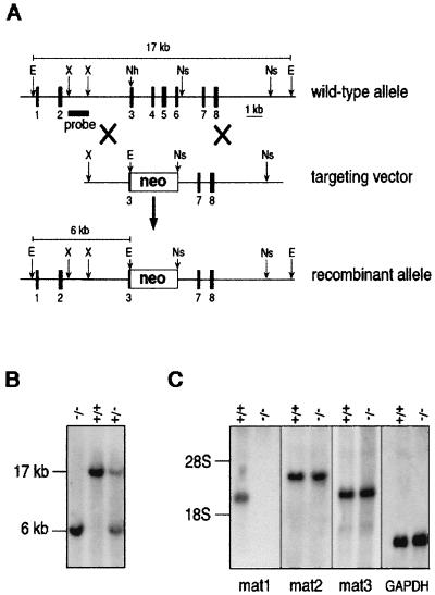
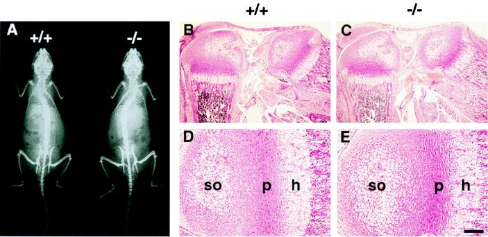
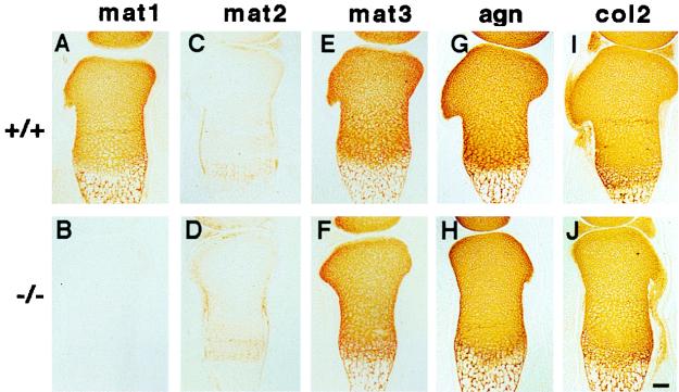
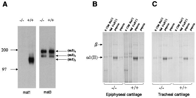
Matrilin 1, or cartilage matrix protein, is a member of a novel family of extracellular matrix proteins. To date, four members of the family have been identified, but their biological role is unknown. Matrilin 1 and matrilin 3 are expressed in cartilage, while matrilin 2 and matrilin 4 are present in many tissues. Here we describe the generation and analysis of mice carrying a null mutation in the Crtm gene encoding matrilin 1. Anatomical and histological studies demonstrated normal development of homozygous mutant mice. Northern blot and biochemical analyses show no compensatory up-regulation of matrilin 2 or 3 in the cartilage of knockout mice. Although matrilin 1 interacts with the collagen II and aggrecan networks of cartilage, suggesting that it may play a role in cartilage tissue organization, studies of collagen extractability indicated that collagen fibril maturation and covalent cross-linking were unaffected by the absence of matrilin 1. Ultrastructural analysis did not reveal any abnormalities of matrix organization. These data suggest that matrilin 1 is not critically required for cartilage structure and function and that matrilin 1 and matrilin 3 may have functionally redundant roles.


Figures

FIG. 1
FIG. 1
Targeted disruption of mouse Crtm encoding matrilin 1. (A) Structure of wild-type allele, targeting construct, and recombinant locus. Dark boxes represent exons of Crtm. The expected fragment sizes after EcoRI digestion are 17 kb for the wild-type allele and 6 kb for the recombinant allele. E, EcoRI; Nh, NheI; Ns, NsiI; X, XhoI. (B) Southern blot analysis of mouse tail DNA isolated from the progeny of a mating between heterozygous parents. DNAs were digested with EcoRI and hybridized with the probe indicated in panel A. +/+, wild-type mouse; +/−, heterozygous mouse; −/−, homozygous mutant mouse. (C) Northern blot analysis of total RNA from limb cartilage, derived from 3-day-old wild-type (+/+) and homozygous mutant mice. The same filter was hybridized with cDNA probes specific for matrilin 1 (mat1), matrilin 2 (mat2), matrilin 3 (mat3), and glyceraldehyde phosphodehydrogenase (GAPDH).
FIG. 2
FIG. 2
Analysis of the skeleton in wild-type (+/+) and matrilin 1-deficient (−/−) mice. (A) X-ray of an 8-week-old mutant mouse shows no gross skeletal abnormalities compared to a normal littermate. (B to E) Hematoxylin-eosin staining of knee region (B and C) and tibia (D and E) from 3-day-old wild-type (B and D) and homozygous mutant (C and E) mice. h, hypertophic zone; p, proliferative zone; so, center of secondary ossification. Bars, 200 μm for panels B and C and 50 μm for panels D and E.
FIG. 3
FIG. 3
Immunostaining of cartilage. Consecutive sections of the tibia from wild-type (+/+ [A, C, E, G, and I]) and matrilin 1-deficient (−/− [B, D, F, H, and J]) newborn littermates were stained with specific antibodies against matrilin 1 (mat1), matrilin 2 (mat2), matrilin 3 (mat3), aggrecan (agn), and type II collagen (col2). Bar, 100 μm.
FIG. 4
FIG. 4
Biochemical analysis of wild type (+/+) and mutant (−/−) epiphyseal and tracheal cartilages. (A) Western blot analysis of matrilin 1 (mat1) and matrilin 3 (mat3) expression in pooled 4 M GuHCl extracts from 3-day-old mouse epiphyseal cartilage. The identities of the matrilin 1 trimers (m1)3 and matrilin 3 trimers (m3)3 and tetramers (m3)4 were determined by comparison with the migration position of protein molecular mass markers (kilodaltons). (B and C) Coomassie-stained SDS–5% polyacrylamide gel of collagens sequentially extracted with neutral salt (0.15 M NaCl), 4 M GuHCl, and pepsin from pooled 7-day-old mouse epiphyseal (B) and tracheal (C) cartilages. The identities of the cartilage collagen α1(II) band and the covalently cross-linked collagen β-components are indicated.

References

    1. Aszódi A, Módis L, Páldi A, Rencendorj A, Kiss I, Bösze Z. The zonal expression of chicken cartilage matrix protein in the developing skeleton of transgenic mice. Matrix Biol. 1994;14:181–190. - PubMed
    1. Aszódi A, Hauser N, Studer D, Paulsson M, Hiripi L, Bösze Z. Cloning, sequencing and expression analysis of mouse cartilage matrix protein cDNA. Eur J Biochem. 1996;236:970–977. - PubMed
    1. Aszódi A, Beier D, Hiripi L, Bösze Z, Fässler R. Sequence, structure and chromosomal localization of Crtm gene encoding mouse cartilage matrix protein and its exclusion as a candidate for murine achondroplasia. Matrix Biol. 1998;16:563–573. - PubMed
    1. Aszódi A, Chan D, Hunziker E, Bateman J F, Fässler R. Collagen II is essential for the removal of the notochord and the formation of intervertebral discs. J Cell Biol. 1998;143:1399–1412. - PMC - PubMed
    1. Bateman J F, Golub S. Deposition and selective degradation of structurally-abnormal type I collagen in an in vitro collagen matrix produced by osteogenesis imperfecta fibroblasts. Matrix Biol. 1994;14:251–262. - PubMed

Publication types

LinkOut - more resources