Comparative Study
. 1999 Nov;19(11):7846-56.
doi: 10.1128/MCB.19.11.7846.

The levels of the bancal product, a Drosophila homologue of vertebrate hnRNP K protein, affect cell proliferation and apoptosis in imaginal disc cells

Affiliations
Comparative Study

The levels of the bancal product, a Drosophila homologue of vertebrate hnRNP K protein, affect cell proliferation and apoptosis in imaginal disc cells

B Charroux et al. Mol Cell Biol. 1999 Nov.

Abstract

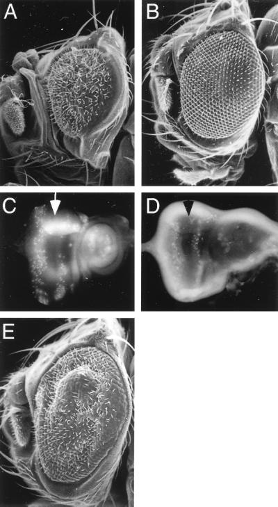
We have characterized the Drosophila bancal gene, which encodes a Drosophila homologue of the vertebrate hnRNP K protein. The bancal gene is essential for the correct size of adult appendages. Reduction of appendage size in bancal mutant flies appears to be due mainly to a reduction in the number of cell divisions in the imaginal discs. Transgenes expressing Drosophila or human hnRNP K are able to rescue weak bancal phenotype, showing the functional similarity of these proteins in vivo. High levels of either human or Drosophila hnRNP K protein in imaginal discs induces programmed cell death. Expression of the antiapoptotic P35 protein suppresses this phenotype in the eye, suggesting that apoptosis is the major cellular defect caused by overexpression of K protein. Finally, the human K protein acts as a negative regulator of bancal gene expression. We propose that negative autoregulation limits the level of Bancal protein produced in vivo.


Figures

FIG. 1
FIG. 1
The reduction in size of adult appendages observed in the blv6/blv4 mutant is rescued by ubiquitous expression of Bl protein. The blv6/blv4 mutant eyes (B), legs (E), and wing (H) are smaller than wild type (A, D, and G). blv6/blv4, hsp70-bl/hsp70-bl flies show normal adult structures when grown at 25°C (C, F, and I). Eyes were observed by SEM (A to C).
FIG. 2
FIG. 2
Molecular analysis of bl. (A) Map of the bl genomic region. Restriction sites are represented by vertical bars, EcoRI sites above and XbaI sites below. Breakpoints or deletions found in bl alleles are indicated by shaded boxes. The location and polarity of the insertion were determined by Southern blot and sequence analysis (data not shown). Genomic λ phages are indicated. The triangle indicates the P-element insertion site. (B) Northern blot analysis revealing bl transcripts, using CR22 cDNA as a probe. mRNAs of 1.9 and 2.1 kb are detected during the first 10 h of embryogenesis (0 to 5 h, lane 1; 5 to 10 h, lane 2); only the larger transcript is detected in the later stages of embryogenesis (lane 3), in third-instar larvae (lane 4), and in adult females (lane 5). Each lane contains 5 μg of poly(A)+ mRNA.
FIG. 3
FIG. 3
The Bl protein is structurally and functionally homologous to human K. (A) Amino acid sequence alignment between Bl and human, Xenopus, and C. elegans K proteins. Identicals amino acids are indicated by black boxes. The Bl protein has a putative nuclear localization signal (NLS) in the N-terminal region and three maxi-KH domains (KH1, KH2, and KH3; in yellow boxes). The KH3 domain is located in the C-terminal extremity of the protein and is separated from the KH2 motif by a hinge region composed of 33% glycine residues for Bl and 19% glycine and 16% proline residues for the human hnRNP K protein. The KNS domain (50) present in the human K protein is indicated. A putative M9 motif, responsible for the shuttling activity of human hnRNP A1 and B2 proteins (49), is present in the Bl protein (amino acids 272 to 319; in blue). (B) Another differentially spliced form of bl transcript produces an abortive protein. Shown is sequence alignment between the bl cDNA CR22 and the EST (expressed sequence tag) clone A1389318. The predicted ORFs of CR22 and the EST clone are indicated. In italics are the amino acids that differs between the two ORFs. The KH1 domain is underlined. (C and D) Binding of the Bl and human K proteins to ribonucleotide homopolymers. The Bl protein and the human K protein were produced by in vitro transcription-translation of pBS-CR22 and pBS-HK5, respectively. An amount equivalent to 20% of the material used for each binding reaction is shown in the lanes marked Translation. The translated proteins were incubated with the indicated ribonucleotide homopolymers in the presence of 200 mM NaCl, and bound proteins were analyzed by SDS-PAGE as described previously (73). Positions of the molecular mass markers are indicated on the left. (E) Amino acid sequence comparison between the M9 motifs identified in different organisms. Conserved residues are underlined. The derived consensus is indicated below the sequence alignment. An amino acid critical for the shuttling activity of the M9 motif is boxed (49).
FIG. 3
FIG. 3
The Bl protein is structurally and functionally homologous to human K. (A) Amino acid sequence alignment between Bl and human, Xenopus, and C. elegans K proteins. Identicals amino acids are indicated by black boxes. The Bl protein has a putative nuclear localization signal (NLS) in the N-terminal region and three maxi-KH domains (KH1, KH2, and KH3; in yellow boxes). The KH3 domain is located in the C-terminal extremity of the protein and is separated from the KH2 motif by a hinge region composed of 33% glycine residues for Bl and 19% glycine and 16% proline residues for the human hnRNP K protein. The KNS domain (50) present in the human K protein is indicated. A putative M9 motif, responsible for the shuttling activity of human hnRNP A1 and B2 proteins (49), is present in the Bl protein (amino acids 272 to 319; in blue). (B) Another differentially spliced form of bl transcript produces an abortive protein. Shown is sequence alignment between the bl cDNA CR22 and the EST (expressed sequence tag) clone A1389318. The predicted ORFs of CR22 and the EST clone are indicated. In italics are the amino acids that differs between the two ORFs. The KH1 domain is underlined. (C and D) Binding of the Bl and human K proteins to ribonucleotide homopolymers. The Bl protein and the human K protein were produced by in vitro transcription-translation of pBS-CR22 and pBS-HK5, respectively. An amount equivalent to 20% of the material used for each binding reaction is shown in the lanes marked Translation. The translated proteins were incubated with the indicated ribonucleotide homopolymers in the presence of 200 mM NaCl, and bound proteins were analyzed by SDS-PAGE as described previously (73). Positions of the molecular mass markers are indicated on the left. (E) Amino acid sequence comparison between the M9 motifs identified in different organisms. Conserved residues are underlined. The derived consensus is indicated below the sequence alignment. An amino acid critical for the shuttling activity of the M9 motif is boxed (49).
FIG. 4
FIG. 4
Analysis of Bl expression. (A and B) The Bl antiserum is specific for the bl gene product, and blv6/blv4 mutant imaginal discs have a reduced amount of the Bl protein. (A) Equal amounts of protein extracted from wild-type ovaries (1), wild-type embryos (2), and wild-type (3) or blv6/blv4 (4) or blv6/blv6 (5) mutant imaginal discs were resolved by SDS-PAGE and transferred to a nitrocellulose membrane. The bl antiserum detects a single 54-kDa protein among proteins prepared from all tissues except proteins prepared from blv6/blv6 (lane 5) mutant imaginal discs. Bl is still detected in blv6/blv4 imaginal discs (lane 4) but at a lower than wild-type level (lane 3). (B) Ponceau S staining of the nitrocellulose membrane immunoblotted by Bl antiserum in panel A. (C to F) Immunodetection of the Bl protein during embryogenesis. (C) Preferential cytoplasmic localization of Bl, syncytial blastoderm, cycle 12. (D) Nuclear and cytoplasmic localization, cellular blastoderm, cycle 13. Cytoplasmic staining is indicated by a thin arrow. Note that the Bl protein is present in the cytoplasm but not in the nucleus of the pole cells at this stage (thick arrow). (E) Nuclear localization, cellular blastoderm, cycle 14. (F) Embryo at stage 9 during germ band elongation. Bl is weakly detected in cells undergoing mitosis (indicated by asterisks) (19); the long arrow indicates the nuclear staining of amnioserosa cells in panel F. In all views, anterior is left and dorsal is uppermost. (C and D) Confocal sections of the posterior region of the embryo; (E and F) optical views.
FIG. 5
FIG. 5
Cell proliferation is reduced in blv6/blv4 mutant imaginal discs. Apoptotic cells were stained with acridine orange in wild-type (A) and blv6/blv4 mutant (B) wing imaginal discs. Arrows in panels A and B indicate acridine orange-positive cells. Inspection of cell size does not reveal any obvious differences between wild-type (C) and blv6/blv4 mutant (D) imaginal discs. Panels D and C correspond to a magnification of the wing blade. (E and F) Imaginal discs from late third instar were labeled with BrdU and stained with anti-BrdU antibodies. The number of fluorescent nuclei reflects the number and distribution of cells in S phase. A significant reduction in the number of BrdU-positive cells is seen in blv6/blv4 third-instar wing discs (F) compared to wild-type discs (E). Arrows indicate the ZNC, and the notum region is boxed. (G and H) Magnifications of the boxed regions in panels E and F, respectively.
FIG. 6
FIG. 6
High levels of hnRNP K proteins induce apoptosis. (A) Immunodetection of the Bl protein in ptcGAL4/UAS-bl wing imaginal disc. The experiment was performed using the anti-Bl antiserum at 1/2,000 dilution to detect only Bl-overexpressing cells. Bl is immunodetected in cells located in a band just anterior (ant) to the anteroposterior boundary. A similar distribution of the human K protein is observed in ptcGAL4/UAS-HK5 wing disc immunostained with mouse monoclonal anti-human K antibody (Fig. 8). post, posterior. (B to D) Wings of wild-type flies (B) or wings of flies overexpressing the Drosophila (C) or the vertebrate (D) K protein under ptcGAL4 regulation lack adult structures. Note the absence of the anterior cross-vein (arrow) and the reduction in size of the intervein tissue between veins 3 and 4 (black dot). These structures originate from the domain that overexpresses the K proteins in imaginal discs (A). (E to G) Apoptotic cells were stained with acridine orange in a wild-type wing imaginal disc (E) and a ptcGAL4/UAS-bl (F) or ptcGAL4/UAS-HK5 (G) wing disc. (H and I) Digoxigenin-labeled rpr cDNA was hybridized to fixed wild-type wing imaginal disc (H) and a ptcGAL4/UAS-bl (I) or a ptcGAL4/UAS-HK5 (J) wing disc. The rpr probe hybridizes to regions that probably overexpress the K proteins.
FIG. 7
FIG. 7
P35 is a suppressor of the phenotype of Bl overexpression in the eye. (A and B) Scanning electron micrographs of GMR-bl2/+ (A) and GMR-P35/+ (B) eyes. (C) High levels of Bl protein in postmitotic cells of the eye lead to increased apoptosis visualized by acridine orange staining (C). (D) Wild-type eye imaginal disc stained with acridine orange. Very little cell death occurs posterior to the morphogenetic furrow (arrows in panels C and D) in a wild-type eye disc. (E) Scanning electron micrograph of a GMR-bl2/+; GMR-P35/+ eye.
FIG. 8
FIG. 8
The human K protein negatively regulates expression of the bl gene. Wing imaginal discs of ptcGAL4/UAS-HK5 third-instar larvae were double stained with anti-human K protein (red) and anti-Bl (green) (A and B), anti-human K protein (red) and anti-Tsh (green) (C), or anti-human K protein (red) and anti-Mod (green) (D). (A and B) Bl is detected in the nuclei of all cells except those expressing the human K protein. Note that like Bl, the human K protein is detected essentially in the nucleus. The arrow in panel B indicates cells expressing both the vertebrate and Drosophila K proteins. Overexpression of the human K protein has no detectable effect on the immunodetection of the Tsh (expressed only in the proximal region of the disc) (C) or Mod (D) protein. Digoxigenin-labeled bl cDNA (E) and mod cDNA (F) were hybridized to fixed ptcGAL4/UAS-HK5 wing imaginal discs. bl transcript is not detected in a band of cells along the anterior-posterior boundary of Gal4-expressing cells (E). Ubiquitous expression of mod transcript is not affected in ptcGAL4/UAS-HK5 wing imaginal discs (F).

References

    1. Alexandre E, Graba Y, Fasano L, Gallet A, Perrin L, De Zulueta P, Pradel J, Kerridge S, Jacq B. The Drosophila Teashirt homeotic protein is a DNA-binding protein and modulo, a HOM C regulated modifier of variegation, is a likely candidate for being a direct target gene. Mech Dev. 1996;59:191–204. - PubMed
    1. Amrein H, Gorman M, Nothiger R. The sex-determining gene tra-2 of Drosophila encodes a putative RNA binding protein. Cell. 1988;55:1025–1035. - PubMed
    1. Baker N E, Rubin G M. Ellipse mutations in the Drosophila homologue of the EGF receptor affect pattern formation, cell division, and cell death in eye imaginal discs. Dev Biol. 1992;150:381–396. - PubMed
    1. Bell L R, Maine E M, Schedl P, Cline T W. Sex-lethal, a Drosophila sex determination switch gene, exhibits sex-specific RNA splicing and sequence similarity to RNA binding proteins. Cell. 1988;55:1037–1046. - PubMed
    1. Bellen H J, Kooyer S, D’Evelyn D, Pearlman J. The Drosophila couch potato protein is expressed in nuclei of peripheral neuronal precursors and shows homology to RNA-binding proteins. Genes Dev. 1992;6:2125–2136. - PubMed

Publication types

MeSH terms

Substances

Associated data

LinkOut - more resources