Comparative Study
. 1999 Nov;19(11):7870-85.
doi: 10.1128/MCB.19.11.7870.

Biological and regulatory properties of Vav-3, a new member of the Vav family of oncoproteins

Affiliations
Comparative Study

Biological and regulatory properties of Vav-3, a new member of the Vav family of oncoproteins

N Movilla et al. Mol Cell Biol. 1999 Nov.

Abstract

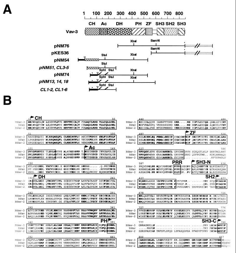
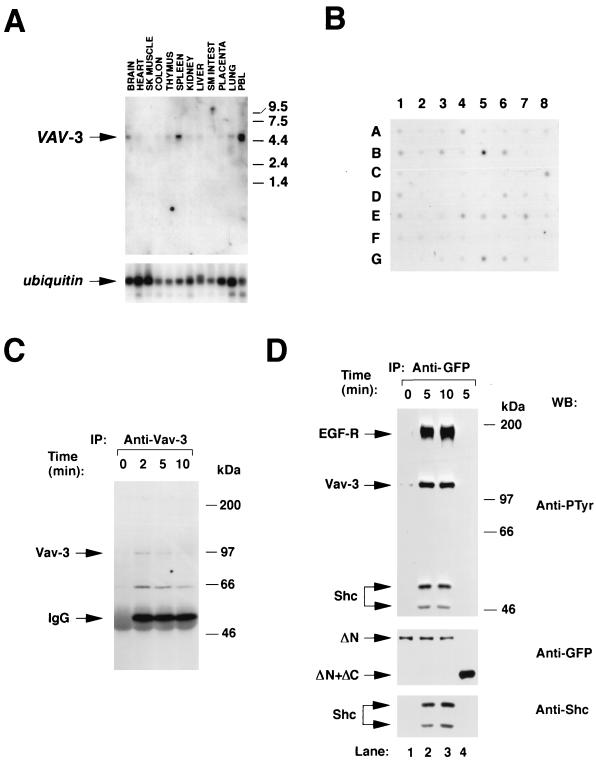
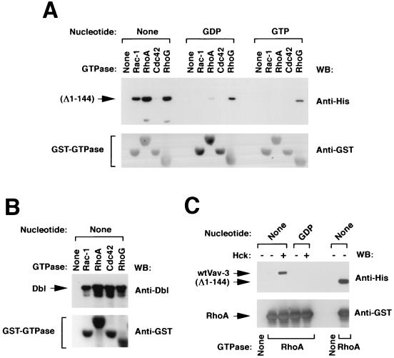
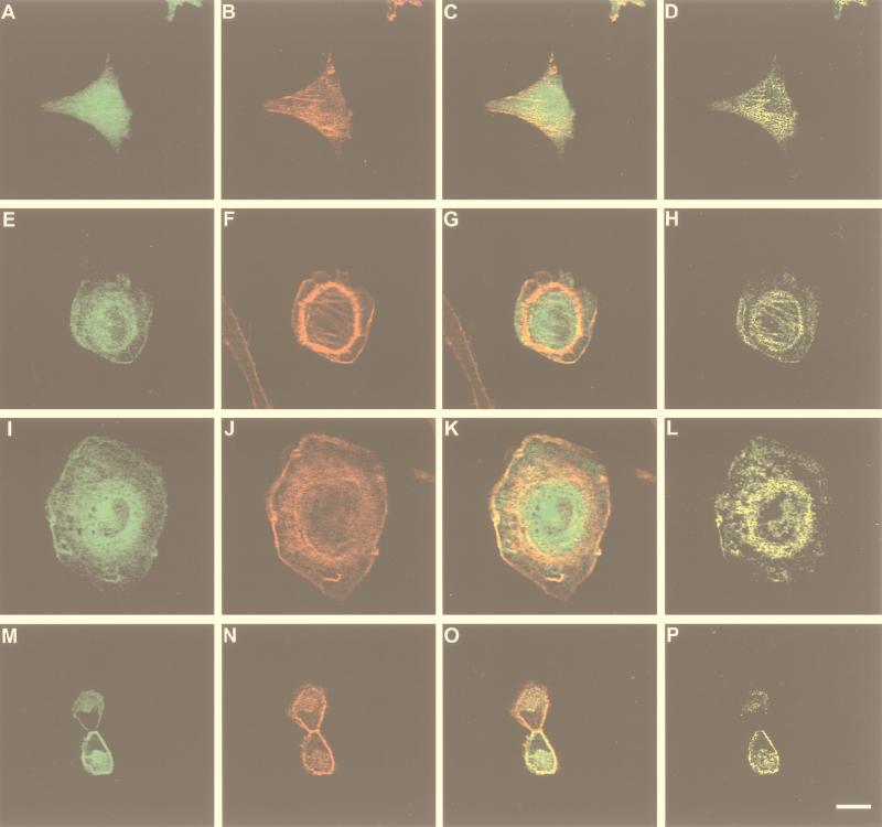
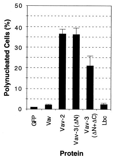
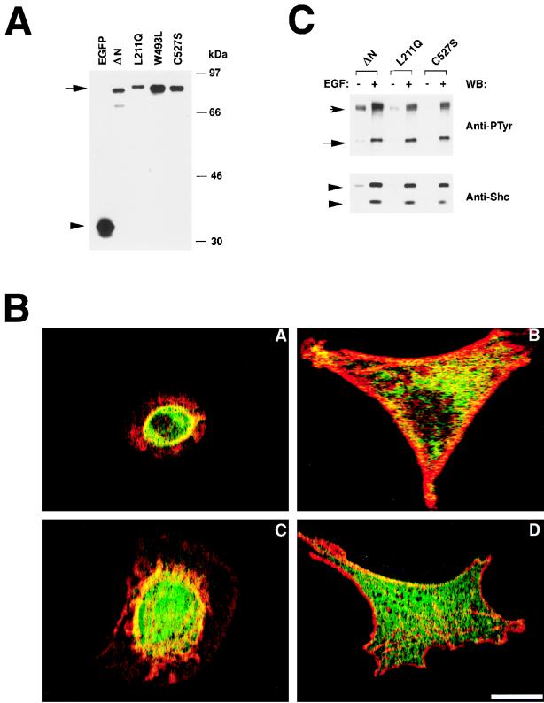
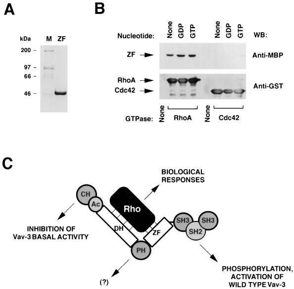
We report here the identification and characterization of a novel Vav family member, Vav-3. Signaling experiments demonstrate that Vav-3 participates in pathways activated by protein tyrosine kinases. Vav-3 promotes the exchange of nucleotides on RhoA, on RhoG and, to a lesser extent, on Rac-1. During this reaction, Vav-3 binds physically to the nucleotide-free states of those GTPases. These functions are stimulated by tyrosine phosphorylation in wild-type Vav-3 and become constitutively activated upon deletion of the entire calponin-homology region. Expression of truncated versions of Vav-3 leads to drastic actin relocalization and to the induction of stress fibers, lamellipodia, and membrane ruffles. Moreover, expression of Vav-3 alters cytokinesis, resulting in the formation of binucleated cells. All of these responses need only the expression of the central region of Vav-3 encompassing the Dbl homology (DH), pleckstrin homology (PH), and zinc finger (ZF) domains but do not require the presence of the C-terminal SH3-SH2-SH3 regions. Studies conducted with Vav-3 proteins containing loss-of-function mutations in the DH, PH, and ZF regions indicate that only the DH and ZF regions are essential for Vav-3 biological activity. Finally, we show that one of the functions of the Vav-3 ZF region is to work coordinately with the catalytic DH region to promote both the binding to GTP-hydrolases and their GDP-GTP nucleotide exchange. These results highlight the role of Vav-3 in signaling and cytoskeletal pathways and identify a novel functional cross-talk between the DH and ZF domains of Vav proteins that is imperative for the binding to, and activation of, Rho GTP-binding proteins.


Figures

FIG. 1
FIG. 1
(A) Schematic representation of the VAV-3 cDNAs isolated in this study. Clones obtained from the screening of cDNA libraries are shown in regular typeface. Those obtained from RACE amplifications are shown in italics. The 5′ UTRs are shown as closed boxes. The 3′ UTRs are shown as open boxes and not in scale. The 5′ ends encoding the Vav-3 CH domain are shown as shaded boxes. Stop codons are indicated by asterisks. Start codons are indicated by inverted triangles. Important restriction sites for cloning purposes are indicated. (B) Alignment of the amino acid sequences of the three members of the Vav family in Homo sapiens. Identical residues present in the three proteins are shown on boldface. The individual structural domains are boxed. PRR, proline-rich region.
FIG. 2
FIG. 2
(A) Distribution of VAV-3 transcripts in adult human tissues. A filter containing 2 μg of poly(A) mRNA isolated from the indicated tissues (MTN blot; Clontech) was hybridized to either a VAV-3 (upper panel) or a ubiquitin (lower panel) probe as described in Materials and Methods. The hybridized filter was exposed for 48 h (upper panel) and 1 h (lower panel). The migration of the VAV-3 and ubiquitin mRNAs is indicated by arrows. The migration of molecular weight markers is indicated on the right. (B) Dot blot analysis of the expression of the VAV-3 gene in adult and embryonic human tissues. The hybridized filter (Human RNA Master blot; Clontech) contained poly(A) mRNA from the following sources: A1 (whole brain, 257 ng), A2 (amigdala, 135 ng), A3 (caudate nucleus, 161 ng), A4 (cerebellum, 109 ng), A5 (cerebral cortex, 99 ng), A6 (frontal lobe, 89 ng), A7 (hippocampus, 118 ng), A8 (medulla oblongata, 124 ng), B1 (occipital lobe, 207 ng), B2 (putamen, 202 ng), B3 (substantia nigra, 132 ng), B4 (temporal lobe, 117 ng), B5 (thalamus, 125 ng), B6 (subthalamic nucleus, 127 ng), B7 (spinal cord, 169 ng), C1 (heart, 410 ng), C2 (aorta, 106 ng), C3 (skeletal muscle, 210 ng), C4 (colon, 177 ng), C5 (bladder, 100 ng), C6 (uterus, 114 ng), C7 (prostate, 190 ng), C8 (stomach, 293 ng), D1 (testis, 213 ng), D2 (ovary, 205 ng), D3 (pancreas, 514 ng), D4 (pituitary gland, 331 ng), D5 (adrenal gland, 349 ng), D6 (thyroid gland, 169 ng), D7 (salivary gland, 393 ng), D8 (mammary gland, 93 ng), E1 (kidney, 213 ng), E2 (liver, 429 ng), E3 (small intestine, 275 ng), E4 (spleen, 175 ng), E5 (thymus, 209 ng), E6 (peripheral blood lymphocytes, 94 ng), E7 (lymph node, 164 ng), E8 (bone marrow, 147 ng), F1 (appendix, 152 ng), F2 (lung, 243 ng), F3 (trachea, 235 ng), F4 (placenta, 361 ng), G1 (fetal brain, 137 ng), G2 (fetal heart, 135 ng), G3 (fetal kidney, 210 ng), G4 (fetal liver, 243 ng), G5 (fetal spleen, 149 ng), G6 (fetal thymus, 104 ng), and G7 (fetal lung, 245 ng). The filter was exposed for 1 week. (C) Phosphorylation of Vav-3 in Jurkat cells. Cells were stimulated for the indicated periods of time, lysed, and immunoprecipitated with antibodies specific for Vav-3. Immunocomplexes were then subjected to anti-phosphotyrosine immunoblot analysis. The mobility of Vav-3 and the immunoglobulin G heavy chain is indicated by arrows. (D) Phosphorylation of Vav-3 proteins in COS-1 cells. Transfected cells were made quiescent and then stimulated for the indicated periods of time with EGF. Proteins were then immunoprecipitated with anti-GFP antibodies and immunoblotted with anti-PTyr antibodies (upper panels). The filter was then blotted with anti-GFP (middle panel) and, after being stripped with SDS at 50°C for 30 min, incubated with anti-Shc antibodies (lower panel). The position of the EGF-R, Vav-3, and Shc isoforms is indicated by arrows. Lanes 1 to 3 contain immunoprecipitated EGFP–Vav-3 (Δ1–144) (ΔN). Lane 4 contains immunoprecipitated EGFP–Vav-3 (Δ1–144+Δ607–847) (ΔN+ΔC). (C and D) The mobility of the coelectrophoresed molecular weight markers is indicated on the right of each panel. IP, immunoprecipitation; WB, Western blot.
FIG. 3
FIG. 3
(A) Coomassie staining of a aliquots of purified Vav-3 (Δ1–144) from Sf9 cells. (B) Aliquots of the purified GST-GTPases from E. coli. (C) GTPase activity of purified Rho proteins estimated as described in Materials and Methods. The mobility of GTP and GDP molecules is indicated. (D) Exchange activity of Vav-3 (Δ1–144) with [3H]GDP release assays. [3H]GDP-loaded GTPases (15 pmol) were incubated with unlabeled GTP either in the absence (black columns) or in the presence (gray columns) of purified Vav-3 (Δ1–144) protein (30 pmol) (left panel). After 45 min at room temperature, aliquots from each incubation were taken in duplicates, and the exchange obtained under each experimental condition was determined by a filter immobilization assay. A similar incubation was performed for 30 min with lysates from either mock (black columns)- or Dbl (light columns)-infected Sf9 cells (right panel). (E) Exchange activity of Vav-3 (Δ1–144) by [35S]GTP-γS incorporation assays. The indicated GDP-loaded GTPases (4 pmol) were incubated with [35S]GTP-γS in the absence (black columns) or presence (gray columns) of purified Vav-3 (Δ1–144) (1.8 pmol). Exchange values were determined as in panel A. In panels D and E, values represent the mean and standard deviation (SD) of three independent determinations each performed in duplicate.
FIG. 4
FIG. 4
(A) Coomassie staining of aliquots of wild-type Vav-3 purified from Sf9 cells. (B) GDP-loaded RhoA (4 pmol) was incubated with [35S]GTP-γS in the presence of 2.0 pmol of either nonphosphorylated Vav-3 (□), nonphosphorylated Vav-3 (Δ1–144) (○), phosphorylated Vav-3 (■), or phosphorylated Vav-3 (Δ1–144) (●). As a negative control, RhoA was also incubated with GST-Lck (◊). Exchange values were determined at the indicated time points as indicated in Fig. 3D. (C) Exchange activity of Vav-3 on Rho/Rac family members by using either 3:1 (left panel) or 1:3 (right panel) molar ratios of GEF-GTPase. The indicated GDP-loaded GTPases were incubated for 45 min at room temperature with [35S]GTP-γS in the presence of GST-Lck (open boxes), nonphosphorylated Vav-3 (gray boxes), or Lck-phosphorylated Vav-3 (closed boxes). Exchange activities were determined as in Fig. 3D. (D) Phosphorylation levels of Vav-3 proteins. Purified wild-type Vav-3 (WT) and Vav-3 (Δ1–144) were separated electrophoretically and subjected to immunoblot analysis with anti-hexahistidine (upper panel) and anti-phosphotyrosine (lower panel) antibodies.
FIG. 5
FIG. 5
(A) Binding of Vav-3 (Δ1–144) to Rho family proteins. Vav-3 (Δ1–144) was incubated with the indicated GTPases as described in Materials and Methods. Bound proteins were separated by SDS-PAGE and subjected to immunoblot analysis by using either anti-hexahistidine residues (upper panel) or anti-GST (lower panel) antibodies. The type of guanosine nucleotide bound to the GTPases used in this study is indicated at the top. (B) Binding of Dbl to Rho proteins. Total cellular lysates from baculovirus-infected Sf9 cells were incubated with the indicated GTPases and processed as indicated above. Blots were incubated with anti-Dbl antibodies and then with anti-GST antibodies to visualize the appropriate proteins. (C) Binding of wild type Vav-3 to RhoA. Wild-type Vav-3 and Vav-3 (Δ1–144) were incubated in kinase buffer alone (−) or with Hck (+) for 15 min and then incubated with either beads alone (None) or coated with GST-RhoA in the indicated nucleotide states. Binding of Vav-3 proteins to RhoA in each experimental condition was determined by immunoblot analysis with anti-hexahistidine antibodies. The mobility of Vav-3 and Vav-3 (Δ1–144) is indicated by arrows. The nucleotide state of each RhoA protein is indicated at the top. In panels A to C the blotting antibody is indicated on the right.
FIG. 6
FIG. 6
Biological effect and subcellular distribution of Vav-3 in transiently transfected NIH 3T3 cells. Cells expressing EGFP (A to D), EGFP–Vav-3 (Δ144–606) (E to H), or EGFP–Vav-3 (Δ1–144+Δ607–847) (I to P) were fixed, stained with phalloidin-rhodamine, and analyzed by confocal microscopy by using filters for fluorescein (A, E, I, and M) or rhodamine (B, F, J, and N). Panels C, G, K, and O show the overlap of both images with the areas of coexpression shown in yellow. Panels D, H, L, and P show only the areas of overlap between Vav-3 and F-actin. The scale bar in panel P is equivalent to 20 μm.
FIG. 7
FIG. 7
Vav-3 induces polynucleated cells. NIH 3T3 cells were transiently transfected with constructs encoding EGFP–Vav-3 (Δ1–144) (Vav-ΔN) and EGFP–Vav-3 (Δ1–144+Δ607–847) (Vav-3ΔN+ΔC) (2 μg each) or with the pEGFP-C1 vector alone (0.25 μg) or in combination with vav-, vav-2-, and LBC-containing plasmids (3 μg each). After 28 h, cells were fixed and then stained with Hoechst 33258 (Sigma), and the number of nuclei present in EGFP-containing cells was scored. Values represent the mean and SD of three independent determinations in which more than 180 cells were counted.
FIG. 8
FIG. 8
(A) Expression of the EGFP–Vav-3 proteins in COS-1 cells. Total cellular lysates from COS-1 cells expressing either EGFP or the indicated EGFP–Vav-3 (Δ1–144+Δ607–847) proteins were subjected to immunoblot analysis with anti-EGFP antibodies. The mobility of Vav-3 proteins is indicated by an arrow. The position of nonchimeric EGFP is marked by an arrowhead. ΔN refers to the Vav-3 (Δ1–144+Δ607–847) protein. (B) Biological activity of Vav-3 proteins. Cells expressing either EGFP–Vav-3 (Δ1–144+Δ607–847) (panel A), EGFP–Vav-3 (Δ1–144+Δ607–847+L211Q) (panel B), EGFP–Vav-3 (Δ1–144+Δ607–847+W293L) (panel C), or EGFP-Vav-3 (Δ1–144+Δ607–847+C527S) (panel D) were fixed, stained with phalloidin-rhodamine, and analyzed by confocal microscopy by using filters for fluorescein (EGFP, green) and rhodamine (actin, red) as indicated in Fig. 6. The panels show the overlap of both images, with the areas of coexpression shown in yellow. In each case, a representative cell of each transfection is shown. Similar results were obtained in three independent transfections. The scale bar in panel D is equivalent to 20 μm. (C) Participation of Vav-3 mutant proteins in the EGF-activated pathway. EGFP–Vav-3 (Δ1–144) (ΔN), Vav-3 (Δ1–144+L211Q) (L211Q), and Vav-3 (Δ1–144+C527S) (C527S) were immunoprecipitated from quiescent (−) and EGF-stimulated (+) COS-1 cells and then tested for phosphorylation (upper panel) and binding to the EGF-R (upper panel) and Shc (lower panel) by using appropriate antibodies. The position of the EGF-R and Vav proteins is indicated by arrows. The position of Shc isoforms is indicated by arrowheads. The blotting antibody is indicated on the right.
FIG. 9
FIG. 9
(A) Schematic representation of the proteins used in this study. The polyhistidine tag is depicted as an open box (not in scale). (B) Vav-3 proteins purified from Sf9 cells. (C) (Left panel). Exchange activity of Vav-3 proteins. Equal molar amounts of each Vav-3 protein were incubated with GDP-loaded RhoA in the presence of free [35S]GTP-γS. After 45 min, aliquots of each reaction were taken in duplicate and the exchange determined as indicated in Fig. 3. Similar results were obtained in two independent experiments, each performed in duplicate. (Right panel) Kinetics of GDP-GTP exchange of RhoA either alone (○) or with Vav-3 (Δ1–144) (□) and Vav-3 (Δ1–144+W293L) (▵). (D) Binding of the indicated Vav-3 proteins to RhoA (upper panel) and immunoblot analysis of the same filter with anti-GST antibodies (lower panel). Experiments were conducted as indicated in Fig. 5. The mobility of Vav-3 (DN) and GST-RhoA is indicated by arrows. The blotting antibody is indicated on the right.
FIG. 10
FIG. 10
(A) Coomassie blue staining of an aliquot of the MBP–Vav-3 ZF protein purified from E. coli cells. (B) The MBP–Vav-3 ZF protein was incubated with the indicated GST-GTPases as described in Materials and Methods. As negative control, MBP–Vav-3 ZF was incubated with beads alone (None). Bound proteins were separated by SDS-PAGE and subjected to immunoblot analysis by using either anti-MBP (upper panel) or anti-GST (lower panel) antibodies. The GTPases used in the study and their nucleotide state are indicated at the bottom and top, respectively. The mobility of the Vav-3 ZF (upper panel) and GTPases (lower panel) is indicated by arrows. The blotting antibody is indicated on the right. (C) Our proposed model for Vav-3 activity.

References

    1. Boguski M S, McCormick F. Proteins regulating Ras and its relatives. Nature. 1993;366:643–654. - PubMed
    1. Bustelo X R. The VAV family of signal transduction molecules. Crit Rev Oncog. 1996;7:65–88. - PubMed
    1. Bustelo X R, Suen K L, Michael W M, Dreyfuss G, Barbacid M. Association of the vav proto-oncogene product with poly(rC)-specific RNA-binding proteins. Mol Cell Biol. 1995;15:1324–1332. - PMC - PubMed
    1. Cerione R A, Zheng Y. The Dbl family of oncogenes. Curr Opin Cell Biol. 1996;8:216–222. - PubMed
    1. Coppola J, Bryant S, Koda T, Conway D, Barbacid M. Mechanism of activation of the vav protooncogene. Cell Growth Differ. 1991;2:95–105. - PubMed

Publication types

MeSH terms

Associated data