

Review
doi: 10.1172/JCI8204.
Th2 cells and GATA-3 in asthma: new insights into the regulation of airway inflammation
Affiliations
- PMID: 10525032
- PMCID: PMC408864
- DOI: 10.1172/JCI8204
Item in Clipboard
Review
Th2 cells and GATA-3 in asthma: new insights into the regulation of airway inflammation
J Clin Invest.
1999 Oct.
No abstract available
Figures
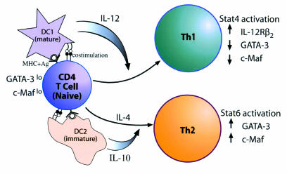
Generation of Th1 and Th2 cells from a naive CD4+ T cell. A naive CD4+ T cell does not secrete cytokines, and has low levels of GATA-3 and c-Maf expression. Differentiation along the Th1 or Th2 pathway is triggered by stimulation with antigen (Ag), presented to the T-cell receptor in the context of MHC by the appropriate APC, and by a second signal, imparted by ligation of costimulatory molecules B7-1/B7-2 and CD28. In the lung, DCs represent the key APC. The DCs have been classified into 2 subsets: mature (DC1) and immature (DC2). The mature DCs express high levels of MHC class II on their surface and produce IL-12, which drives Th1 differentiation. The immature DCs express low levels of MHC class II and produce IL-10, which favors Th2 differentiation. Furthermore, the cytokine(s) present in the microenvironment, IL-4/IL-10 versus IL-12, plays a decisive role in orchestrating the differentiation along the Th1 or Th2 lineage.
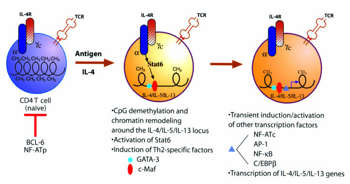
Molecular mechanisms of differentiation of a naive CD4+ T cell into Th2 cells. A naive CD4+ T cell contains a condensed chromatin structure with extensive methylation. Antigen stimulation and engagement of the IL-4R result in STAT6 activation, which, in turn, causes specific demethylation around the IL-4/IL-5/IL-13 locus (similarly, antigen + IL-12 causes demethylation around the IFN-γ locus). Chromatin remodeling is accompanied by induction of Th2-specific transcription factors such as GATA-3 and c-Maf, which bind to target sequences in the IL-4/IL-5/IL-13 locus. The chromatin — rendered accessible by demethylation and perhaps by binding of GATA-3, c-Maf, and other, currently undiscovered Th2-specific transcription factors — is next bound by more widely expressed and transiently induced transcription factors such as AP-1, NF-κB, NF-ATc, and C/EBPβ. This may allow synergistic interactions between the tissue-specific and general transcription factors to occur, resulting in the active transcription of the IL-4, IL-5, and IL-13 genes. Effector/memory cells are thought to be in a state of suspended animation, with an open chromatin structure and high levels of GATA-3 and c-Maf expression. Restimulation of these cells by antigen would result in transient induction of the general factors leading to rapid induction of Th2 gene expression.
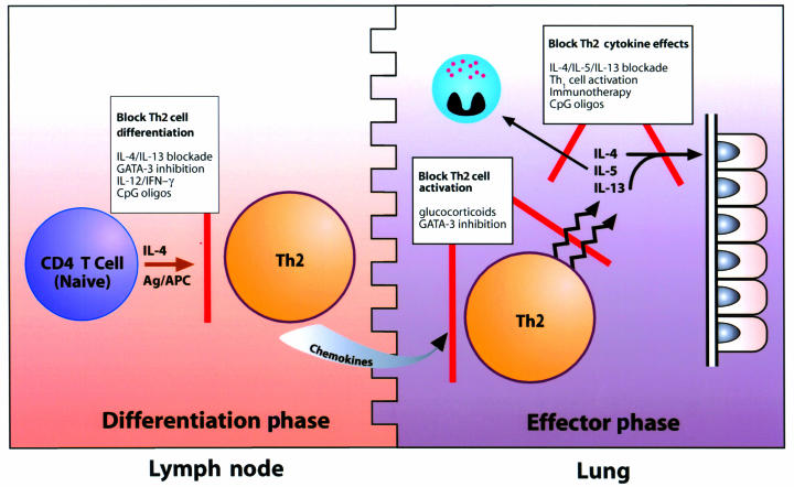
Potential strategies for inhibiting Th2 function in asthma. (a) Block Th2 cell differentiation. The cytokine environment that is present during CD4+ T-cell differentiation affects the development of new Th2 cells. Blocking IL-4 and/or IL-13 or the Th2-specific transcription factor GATA-3 leads to inhibition of Th2 cell induction. Alternatively, increasing levels of IL-12 and IFN-γ in the environment may shift the Th1/Th2 balance toward Th1 and reduce the number of Th2 cells in the respiratory tract. This can be accomplished by stimulating a Th1 response to certain infectious agents, such as Mycobacterium, or by the administration of CpG oligodeoxynucleotides. All of these interventions may have an effect in asthma by reducing the number of Th2 cells in the airways and by inhibiting the generation of new Th2 cells. (b) Block Th2 cell activation. Glucocorticoids are standard therapy in asthma and cause nonselective immunosuppression. Th2 cell activation may be selectively blocked using inhibitors of GATA-3, a T cell–specific factor that controls the production of key Th2 cytokines. (c) Block Th2 cytokine effects. Blocking IL-4, IL-5, or IL-13 may inhibit the effects of these cytokines on target tissues. In addition, Th1 cells have been shown to inhibit Th2 cytokine effects on eosinophils and airway epithelial mucus production. Allergy immunotherapy and CpG oligodeoxynucleotides both induce Th1 cells and lead to a reduction in allergic airway pathology.
References
-
- Wills-Karp M. Immunologic basis of antigen-induced airway induced hyperresponsiveness. Annu Rev Immunol. 1999;17:255–281. - PubMed
-
- Cohn L, et al. Th2-induced airway mucus production is dependent on IL-4Ralpha, but not on eosinophils. J Immunol. 1999;162:6178–6183. - PubMed
-
- Corrigan CJ, Hartnell A, Kay AB. T lymphocyte activation in acute severe asthma. Lancet. 1988;1:1129–1132. - PubMed
-
- Walker C, et al. Allergic and nonallergic asthmatics have distinct patterns of T-cell activation and cytokine production in peripheral blood and bronchoalveolar lavage. Am Rev Respir Dis. 1992;146:109–115. - PubMed
Publication types
MeSH terms
Substances
Grants and funding
LinkOut - more resources
Full Text Sources
Other Literature Sources
Medical
Research Materials
Miscellaneous
