. 1999 Oct;104(8):1061-9.
doi: 10.1172/JCI6685.

Expression of functional CXCR4 chemokine receptors on human colonic epithelial cells

Affiliations

Expression of functional CXCR4 chemokine receptors on human colonic epithelial cells

N J Jordan et al. J Clin Invest. 1999 Oct.

Abstract

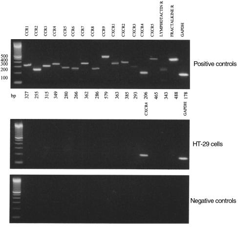
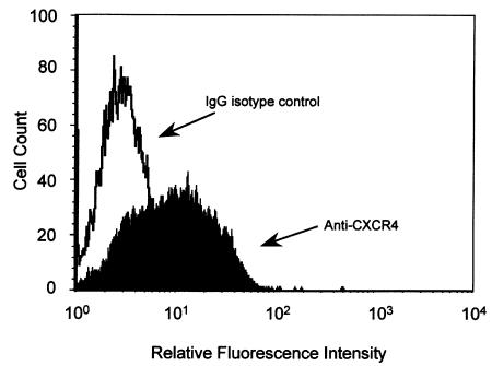
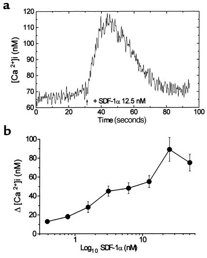
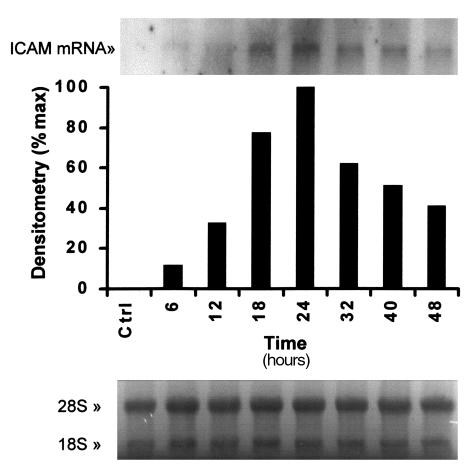
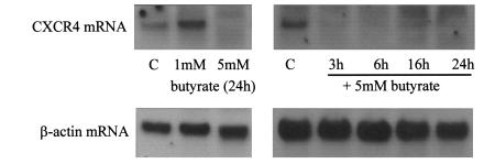
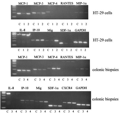
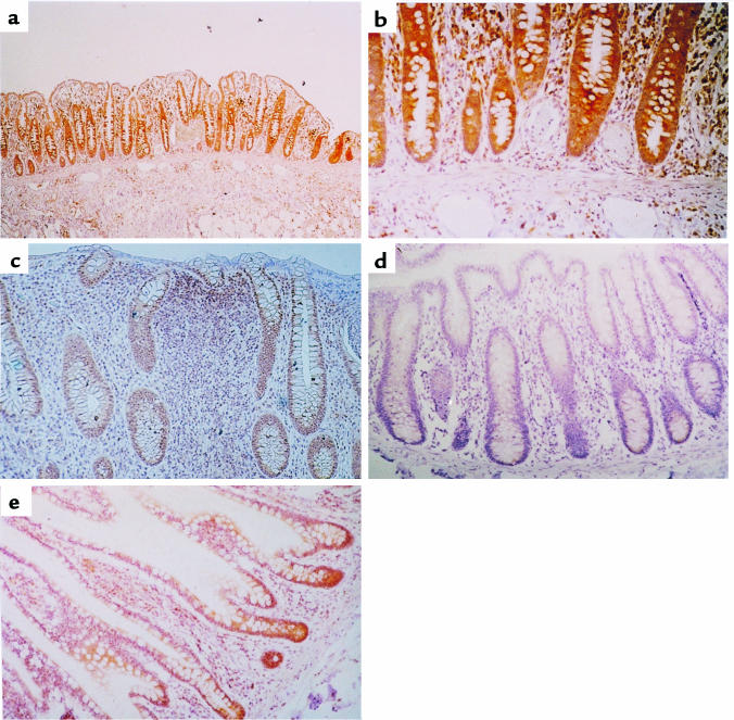
In addition to their role as regulators of leukocyte migration and activation, chemokines and their receptors also function in angiogenesis, growth regulation, and HIV-1 pathogenesis--effects that involve the action of chemokines on nonhematopoietic cells. To determine whether chemokine receptors are expressed in human colonic epithelium, HT-29 cells were examined by RT-PCR for the expression of the chemokine receptors for lymphotactin, fractalkine, CCR1-10, and CXCR1-5. The only receptor consistently detected was CXCR4 (fusin/LESTR), although HT-29 cells did not express mRNA for its ligand, stromal cell-derived factor (SDF-1alpha). Flow cytometric analysis with anti-CXCR4 antibody indicated that the CXCR4 protein was expressed on the surface of roughly half of HT-29 cells. CXCR4 was also expressed in colonic epithelial cells in vivo as shown by immunohistochemistry on biopsies from normal and inflamed human colonic mucosa. The mRNA for SDF-1alpha and other CC and CXC chemokines was present in normal colonic biopsies. The CXCR4 receptor in HT-29 cells was functionally coupled, as demonstrated by the elevation in [Ca2+]i, which occurred in response to 25 nM SDF-1alpha and by the SDF-1alpha-induced upregulation of ICAM-1 mRNA. Sodium butyrate downregulated CXCR4 expression and induced differentiation of HT-29 cells, suggesting a role for CXCR4 in maintenance and renewal of the colonic epithelium. This receptor, which also serves as a coreceptor for HIV, may mediate viral infection of colonic epithelial cells.


Figures

Figure 1
Figure 1
Expression of chemokine receptor mRNA in HT-29 cells. PCR analysis was performed using primers against the chemokine receptors CCR1-9/10 and CXCR1-5 with GAPDH as a housekeeping probe as described in Methods. A mixed leukocyte preparation (or Jurkats) was used as a positive control, as shown in the top panel. HT-29 cells expressed CXCR4, as shown in the middle panel, and the negative control (HT-29 mRNA, which was not reverse transcribed) was consistently negative, as shown in the bottom panel. The experiment shown is representative of at least 3 independent experiments.
Figure 2
Figure 2
Surface expression of CXCR4 on HT-29 cells. HT-29 cells were incubated with 10 μg/mL anti-CXCR4 (filled) or an isotype control antibody (open), then labeled with a FITC conjugate. Washed cells were analyzed by flow cytometry in which accumulated events were gated against the isotype control. The experiment shown is representative of 3 similar experiments.
Figure 3
Figure 3
Ca2+ flux in HT-29 cells in response to SDF-1α. (a) The elevation of intracellular Ca2+ induced in fura-2/AM–loaded HT-29 cells in response to stimulation with 12.5 nM SDF-1α. (b) The response to SDF-1α is concentration-dependent. Aliquots of fura-2/AM–loaded HT-29 cells were stimulated with SDF-1α at concentrations from 0.4 to 50 nM. Results are the mean [Ca2+]i for 3–14 measurements (± SEM).
Figure 4
Figure 4
SDF-1α enhances ICAM-1 mRNA expression in HT-29 cells. HT-29 cells were serum starved for 24 hours, then treated between 1 and 48 hours with 25 nM SDF-1α, before RNA was extracted. Blots were probed for ICAM-1 mRNA. Densitometric analysis shows maximum expression at 24 hours. Equal loading is shown by the ethidium bromide staining of the 18S and 28S ribosomal RNA (bottom panel). The result shown is representative of 2 similar experiments.
Figure 5
Figure 5
CXCR4 mRNA expression in HT-29 cells is reduced by sodium butyrate. HT-29 cells were serum starved for 24 hours and then stimulated with 1 mM or 5 mM sodium butyrate as shown for 3–24 hours. RNA was extracted as described in Methods and analyzed by Northern blotting. Blots were probed for CXCR4 mRNA (top panel) and β-actin to demonstrate equal loading. The result shown is representative of 3 similar experiments.
Figure 6
Figure 6
Chemokine expression in HT-29 cells and human colon. The top 2 panels show the expression of chemokine mRNA by colonic epithelial cells using RT-PCR analysis of treated HT-29 cells. HT-29 cells were serum starved for 24 hours, then either left untreated for an additional 6 hours (denoted by the number 1) or treated for 6 hours with IL-1α, TNF-α, and IFN-γ (3 ng/mL, 30 ng/mL, 100 U/mL, respectively) (denoted by the number 2). The mRNA was prepared and PCR analysis performed as described in Methods. A mixed leukocyte preparation was used as a positive control (C) for all primer pairs except SDF-1α, where human bone marrow stromal cells were used as a positive control. The bottom 2 panels show RT-PCR analysis of chemokine mRNA expression in human colonic biopsies taken from 2 patients with normal noninflamed colon (denoted by the numbers 3 and 4).
Figure 7
Figure 7
Immunohistochemistry of human colonic biopsy. Specimens of human colonic mucosa were studied immunohistochemically for the presence of CXCR4 by staining with the anti-CXCR4 antibody 12G5. (a) CXCR4 expression in normal colonic epithelium, endothelial cells, and cells in the lamina propria (×10). (b) Normal colonic epithelium. Staining due to CXCR4 expression is more intense in the cells at the base of the crypt (×40). (c) Ulcerative colitis with CXCR4 expression in colonic epithelial cells of the crypt and cells infiltrating the lamina propria (×25). (d) No staining detected in colonic epithelial cells using an isotype-matched antibody (×25). (e) CXCR4 expression in epithelial cells in the terminal ileum (×25).

Similar articles

Cited by

References

    1. Warhurst AC, Hopkins SJ, Warhurst G. Interferon gamma induces differential upregulation of α and β chemokine secretion in colonic epithelial cell lines. Gut. 1998;42:208–213. - PMC - PubMed
    1. Yang SK, Eckmann L, Panja A, Kagnoff MF. Differential and regulated expression of CXC, CC and C chemokines by human colon epithelial cells. Gastroenterology. 1997;113:1214–1223. - PubMed
    1. Kennedy J, et al. Molecular cloning and functional characterisation of human lymphotactin. J Immunol. 1995;155:203–209. - PubMed
    1. Bazan JF, et al. A new class of membrane-bound chemokine with a CX3C motif. Nature. 1997;385:640–644. - PubMed
    1. Keane MP, Arenberg DA, Moore BB, Addison CL, Streiter RM. CXC chemokines and angiogenesis/angiostasis. Proc Natl Acad Sci USA. 1998;110:288–296. - PubMed

Publication types