. 1999 Oct;104(8):1115-21.
doi: 10.1172/JCI7696.

Mutations in Igalpha (CD79a) result in a complete block in B-cell development

Affiliations

Mutations in Igalpha (CD79a) result in a complete block in B-cell development

Y Minegishi et al. J Clin Invest. 1999 Oct.

Abstract

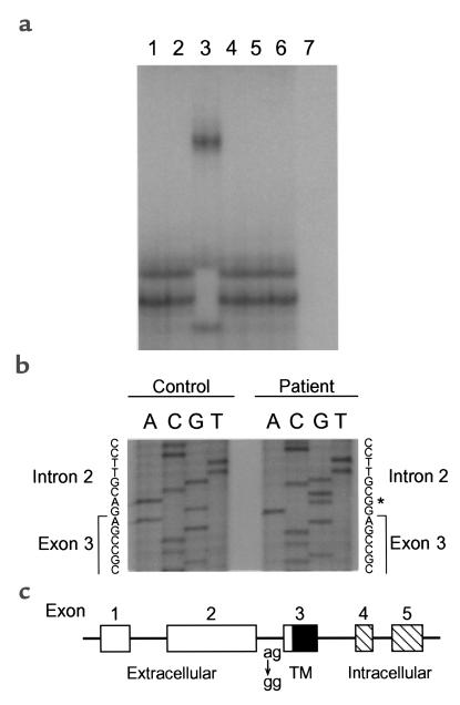
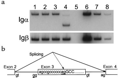
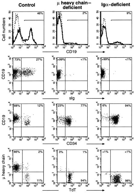
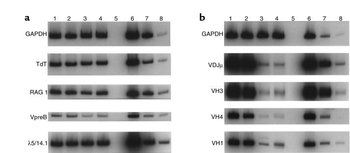
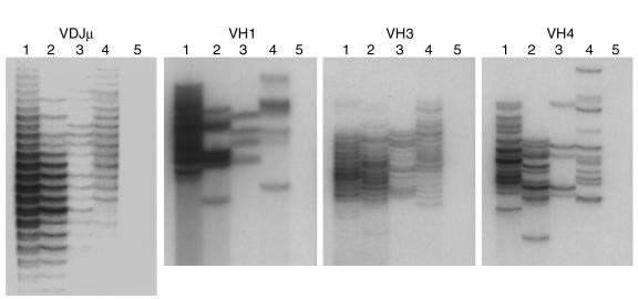
Mutations in Btk, mu heavy chain, or the surrogate light chain account for 85-90% of patients with early onset hypogammaglobulinemia and absent B cells. The nature of the defect in the remaining patients is unknown. We screened 25 such patients for mutations in genes encoding components of the pre-B-cell receptor (pre-BCR) complex. A 2-year-old girl was found to have a homozygous splice defect in Igalpha, a transmembrane protein that forms part of the Igalpha/Igbeta signal-transduction module of the pre-BCR. Studies in mice suggest that the Igbeta component of the pre-BCR influences V-DJ rearrangement before cell-surface expression of mu heavy chain. To determine whether Igalpha plays a similar role, we compared B-cell development in an Igalpha-deficient patient with that seen in a mu heavy chain-deficient patient. By immunofluorescence, both patients had a complete block in B-cell development at the pro-B to pre-B transition; both patients also had an equivalent number and diversity of rearranged V-DJ sequences. These results indicate that mutations in Igalpha can be a cause of agammaglobulinemia. Furthermore, they suggest that Igalpha does not play a critical role in B-cell development until it is expressed, along with mu heavy chain, as part of the pre-BCR.


Figures

Figure 1
Figure 1
Identification of a mutation in the Igα gene. (a SSCP analysis of genomic DNA from a control (lane 1), patients with agammaglobulinemia (lanes 2–6), or no DNA template (lane 7) using primers specific for exon 3 of Igα. The DNA from the patient is shown in lane 3. (b) Sequence analysis revealed a single-bp substitution (A→G) at the invariant –2 position of the splice acceptor site for exon 3. The patient’s mutation is noted by an asterisk. (c) Schematic diagram of the 5 exons constituting the genomic structure of Igα. The extracellular region (open), the transmembrane region (TM; filled), the intracellular region (hatched), and the position of the point mutation in the patient are indicated.
Figure 2
Figure 2
Effects of the Igα mutation on splicing. (a) Bone marrow–derived cDNA from 2 healthy controls (lanes 1 and 2), a patient with μ heavy chain deficiency (lane 3), and the patient with mutations in Igα (lane 4) was amplified using primers from exon 2 and exon 5 of Igα (top) or primers specific for Igβ (bottom). Ten-fold dilutions of control cDNA (10×, 1×, and 0.1×) are shown in lanes 6–8. Sequencing of the smaller band in lane 4 demonstrated skipping of exon 3 of Igα. The sequence of the fainter band in this lane showed the use of a cryptic splice site within exon 3. (b) Schematic diagram showing exon skipping and activation of the cryptic splice site within exon 3. The coding sequence for exon 3 is shown within the central box. The majority of the Igα transcripts amplified in lane 4 in panel (a) delete exon 3, as indicated by the solid black line. A fraction of the transcripts are derived from the use of the cryptic splice site within exon 3, indicated by the dotted line. The invariant dinucleotides at the splice donor and acceptor sites are shown. The position of the point mutation and the dinucleotide at the cryptic acceptor site are underlined. The intron and the coding sequence that arise from the use of the cryptic splice site are shown in lowercase letters and uppercase letters, respectively.
Figure 3
Figure 3
Flow cytometric analysis of B-cell development. Bone marrow mononuclear cells from a control (left column), μ heavy chain–deficient patient (middle column), and Igα-deficient patient (right column) were stained with antibody to CD19. Both patients showed a marked reduction in the number of CD19+ cells within the lymphoid gate (first row). Less than 1% of the CD19+ cells from either patient were B cells, as defined by coexpression of CD19 and surface immunoglobulin (second row). More than 75% of the B-lineage cells in both patients were positive for CD34 (third row). CD19+ permeabilized cells were stained for cytoplasmic TdT and μ heavy chain. More than 94% of the CD19+ cells from both patients were pro-B cells, as defined by coexpression of CD19 and TdT (fourth row).
Figure 4
Figure 4
Semiquantitative RT-PCR analysis of B cell–specific transcripts in the bone marrow. (a) Equal amounts of cDNA from 2 healthy controls (lanes 1 and 2), a patient with μ heavy chain deficiency (lane 3), the patient with Igα deficiency (lane 4), a cDNA negative template control (lane 5), and three 10-fold dilutions of control cDNA (10×, 1×, and 0.1×) (lanes 6–8) were amplified using primers specific for TdT, RAG1, VpreB, and λ5/14.1. GAPDH was used as a control to demonstrate equal concentrations of cDNA. (b) cDNA samples were analyzed as in a, except that the 10-fold dilutions in lanes 6–8 were 1×, 0.1×, and 0.01× of the control. Primers specific for all VH family members or primers specific for VH3, VH4, or VH1 were paired with a primer form the CH1 domain of μ heavy chain.
Figure 5
Figure 5
Repertoire diversity of V-DJ rearranged μ heavy chain transcripts. RT-PCR products from Figure 4 were separated on a denaturing 6% polyacrylamide (sequencing) gel. The control samples (lanes 1 and 2) were diluted 10-fold compared with the patient samples. PCR products from the μ heavy chain–deficient patient are shown in lane 3, and products from the Igα–deficient patient are shown in lane 4.

Comment in

  • Igalpha: B all that you can B.
    Wang LD, Clark MR. Wang LD, et al. J Clin Invest. 1999 Oct;104(8):1011-2. doi: 10.1172/JCI8510. J Clin Invest. 1999. PMID: 10525036 Free PMC article. No abstract available.

References

    1. Karasuyama H, et al. The expression of Vpre-B/lambda 5 surrogate light chain in early bone marrow precursor B cells of normal and B cell-deficient mutant mice. Cell. 1994;77:133–143. - PubMed
    1. Lassoued K, Illges H, Benlagha K, Cooper MD. Fate of surrogate light chains in B lineage cells. J Exp Med. 1996;183:421–429. - PMC - PubMed
    1. Koyama M, et al. CD79α/CD79β heterodimers are expressed on pro-B cell surfaces without associated μ heavy chain. Int Immunol. 1997;9:1767–1772. - PubMed
    1. Nagata K, et al. The Igα/Igβ heterodimer on μ-negative proB cells is competent for transducing signals to induce early B cell differentiation. Immunity. 1997;7:559–570. - PubMed
    1. Gong S, Nussenzweig MC. Regulation of an early developmental checkpoint in the B cell pathway by Igβ. Science. 1996;272:411–414. - PubMed

Publication types