. 1999 Oct 18;147(2):207-20.
doi: 10.1083/jcb.147.2.207.

RAD51 and DMC1 form mixed complexes associated with mouse meiotic chromosome cores and synaptonemal complexes

Affiliations

RAD51 and DMC1 form mixed complexes associated with mouse meiotic chromosome cores and synaptonemal complexes

M Tarsounas et al. J Cell Biol. .

Abstract

The eukaryotic RecA homologues RAD51 and DMC1 function in homology recognition and formation of joint-molecule recombination intermediates during yeast meiosis. The precise immunolocalization of these two proteins on the meiotic chromosomes of plants and animals has been complicated by their high degree of identity at the amino acid level. With antibodies that have been immunodepleted of cross-reactive epitopes, we demonstrate that RAD51 and DMC1 have identical distribution patterns in extracts of mouse spermatocytes in successive prophase I stages, suggesting coordinate functionality. Immunofluorescence and immunoelectron microscopy with these antibodies demonstrate colocalization of the two proteins on the meiotic chromosome cores at early prophase I. We also show that mouse RAD51 and DMC1 establish protein-protein interactions with each other and with the chromosome core component COR1(SCP3) in a two-hybrid system and in vitro binding analyses. These results suggest that the formation of a multiprotein recombination complex associated with the meiotic chromosome cores is essential for the development and fulfillment of the meiotic recombination process.


Figures

Figure 1
Figure 1
Truncation derivatives of the mouse RAD51 and DMC1 proteins used in two-hybrid and in vitro binding analyses. We divided the 340 amino acids into two regions with different degrees of identity between the two proteins. The black boxes depict nucleotide binding motifs (Story et al. 1993; Habu et al. 1996) located in the RecA homology core.
Figure 2
Figure 2
Thrombin removal of the HA-tag. The pET29aHA vector engineered here was tested for solubility of the target protein expressed and retention of the thrombin digestion site. COR1(SCP3) expressed in this system with an HA-tag at the NH2 terminus, was detected in large amounts in the soluble fraction of the bacterial extract with an anti-HA antibody. The thrombin treatment removed the HA tag, indicating that the expression vector functions according to our design.
Figure 4
Figure 4
Specificity of anti-RAD51 and anti-DMC1 antibodies determined using an immunocytological approach. Anti-RAD51 and anti-DMC1 purified antibodies were each reacted with one and the other of the two proteins, the immune complexes removed by centrifugation, and the resulting antibodies analyzed on spermatocyte chromosome spreads. (A, A′, and B) Anti-DMC1 antibody (green) blocked with RAD51 recognizes the expected number of foci at early stages (A and A′; A′ is an enlargements of the area marked in A), while the same antibody blocked with DMC1 does not produce any green fluorescence signal (B). (C, C′, and D) Similarly, the signal generated with the anti-RAD51 antibody (green) is not affected by reaction with DMC1 protein (C and C′), while RAD51 blocks it completely (D). Note that the green fluorescence is enhanced in B and D to detect any possible signal associated with the chromosomes. Chromosomal cores are stained in red with mouse (A, A′, and B) or rabbit (C, C′, and D) anti-COR1 antibodies.
Figure 3
Figure 3
Specificity of the anti-RAD51 and anti-DMC1 antibodies determined on Western blots of affinity-purified proteins. The anti-DMC1 and anti-RAD51 polyclonal antibodies recognize both proteins on Western blot analyses (shown here for anti-DMC1 in A). To ensure specificity for their own antigen, each of the two antibodies was immunodepleted of components cross-reacting with the homologous protein. This procedure renders an antibody with specificity for its antigen (B and D). Equal amounts of the purified protein were loaded into the gel as shown in the Coomassie-stained gel (C). The second band in the RAD51 doublet in D corresponds to a degradation product.
Figure 5
Figure 5
Colocalization of RAD51 and DMC1 proteins with immunogold EM. DMC1 is visualized with a goat anti–mouse secondary antibody conjugated with 5-nm-gold particles. RAD51 is visualized with a goat anti-rabbit secondary antibody conjugated with 10-nm-gold particles. Centromeres are stained with a human CREST serum visualized here with 15-nm-gold grains. (A) In the leptotene configuration, the grains identify mixed RAD51/DMC1 foci attached to individual chromosome cores. (B) At zygotene, the RAD51/DMC1 foci colocalize with axial associations established between homologous chromosomes, the sites of synapsis initiation. The B′ section is enlarged to point out the differences in size between the gold particles. (C) At pachytene, the RAD51/DMC1 foci localize along the fully synapsed chromosomes.
Figure 7
Figure 7
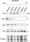
Western blot detection of RAD51, DMC1, and meiotic markers COR1(SCP3) and SYN1(SCP1) in rat spermatocyte fractions isolated by centrifugal elutriation. The composition of each fraction was estimated from the distribution of COR1(SCP3), SYN1(SCP1), and centromeres with immunofluorescence staining. Protein amounts corresponding to equal numbers of cells were loaded into each lane. The presence of COR1(SCP3) and SYN1(SCP1) in this Western blot follows the expected temporal pattern (see Discussion). RAD51 and DMC1 are detected only in fractions enriched in early prophase I spermatocytes, consistent with fluorescence and EM observations.
Figure 6
Figure 6
Colocalization of RAD51 and DMC1 proteins with immunofluorescence labeling. Immunodepleted mouse and rabbit antibodies were used to detect DMC1 and RAD51, respectively, on rat surface-spread spermatocytes. The cores are stained with trace amounts of a mouse anti-COR1(SCP3) antibody, visible in C and F. DNA is stained with DAPI (blue). (A–C) In early prophase I stages (leptotene, L, and zygotene, Z) RAD51 and DMC1 foci are positioned along the cores of homologous chromosomes. To a red-labeled focus in A corresponds a green-labeled focus in B in 95% of the cases. (D, E, and F) At pachytene (P) the RAD51/DMC1 foci are positioned along the SCs and are reduced in number as compared with previous stages.
Figure 8
Figure 8
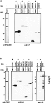
Protein–protein interactions determined in vitro with a pull-down assay. The two His-tagged proteins are detected with specific anti-RAD51 (A) and anti-DMC1 (B) antibodies. The same amount of His-tagged protein bound to a Ni-NTA agarose column was incubated with a soluble bacterial extract expressing the HA-tagged protein (in the amounts shown in the lanes containing HA-tagged protein only). Interacting proteins were coprecipitated. After washes and elution from the beads, the HA-tagged proteins engaged in an interaction with the His-tagged protein were detected with an anti-HA antibody, as described in the Materials and Methods. + Indicates interaction, and − lack of interaction.

References

    1. Albini S.M., Jones G.H. Synaptonemal complex spreading in Allium cepa and A. fistulosum. The initiation and sequence of pairing. Chromosoma. 1987;95:324–338.
    1. Anderson L.K., Stack S.M. Nodules associated with axial cores and synaptonemal complexes during zygotene in Psilotum nudum . Chromosoma. 1988;97:96–100.
    1. Anderson L.K., Offenberg H.H., Verkuijlen W.H.H.C., Heyting C. RecA-like proteins are components of the early meiotic nodules in lily. Proc. Natl. Acad. Sci. USA. 1997;94:6868–6873. - PMC - PubMed
    1. Baker S.M., Plug A.W., Prolla T.A., Bronner C.E., Harris A.C., Yao X., Christie D.M., Monell C., Arnheim N., Bradley A., Ashley T., Liskay R.M. Involvement of mouse MLH1 in DNA mismatch repair and meiotic crossing over. Nat. Genet. 1996;13:336–342. - PubMed
    1. Barlow C., Hirotsune S., Paylor R., Liyanage M., Eckhaus M., Collins F., Shiloh Y., Crawley J.N., Ried T., Tagle D., Wynshaw-Boris A. Atm-deficient micea paradigm of ataxia telangiectasia. Cell. 1996;86:159–171. - PubMed

Publication types