. 1999 Oct 18;147(2):257-66.
doi: 10.1083/jcb.147.2.257.

Glycosylation can influence topogenesis of membrane proteins and reveals dynamic reorientation of nascent polypeptides within the translocon

Affiliations

Glycosylation can influence topogenesis of membrane proteins and reveals dynamic reorientation of nascent polypeptides within the translocon

V Goder et al. J Cell Biol. .

Abstract

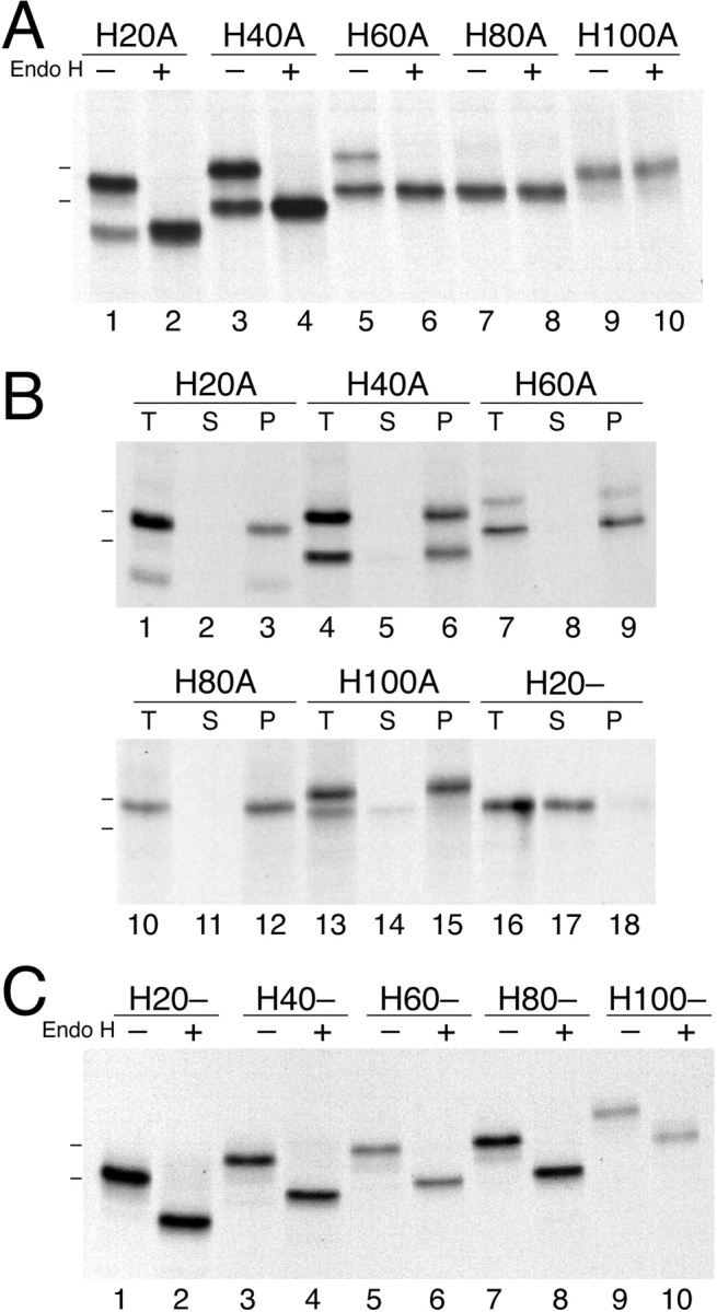
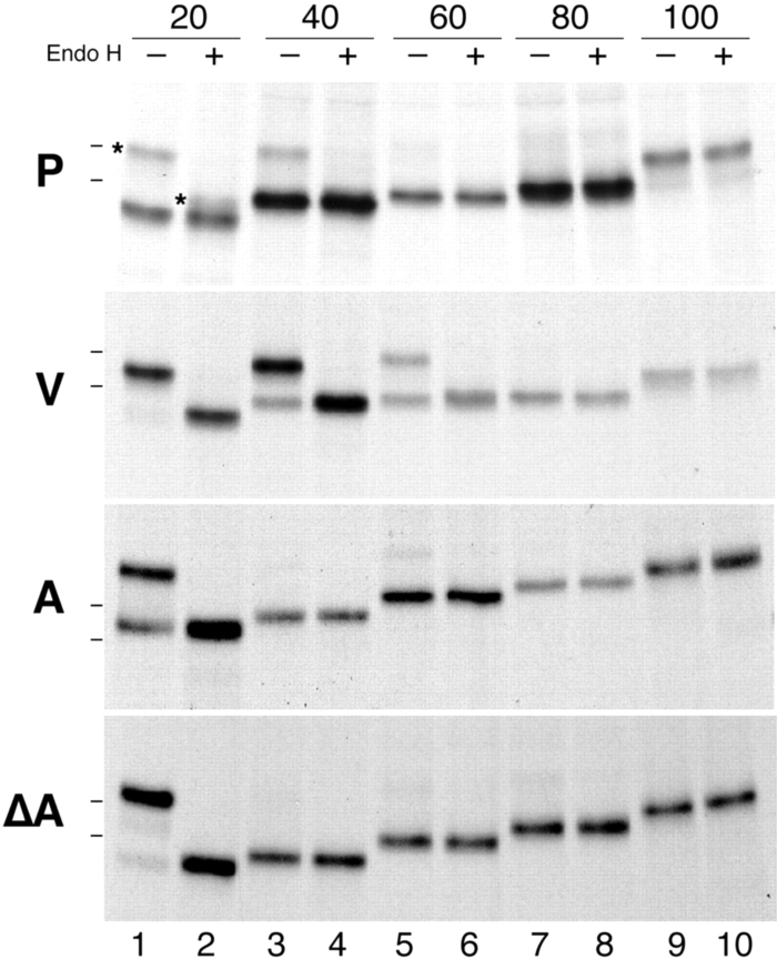
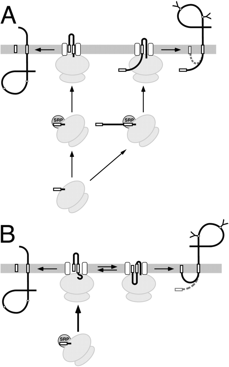
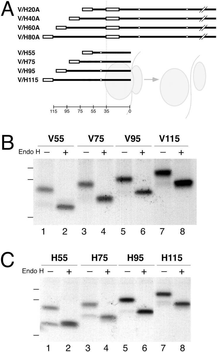
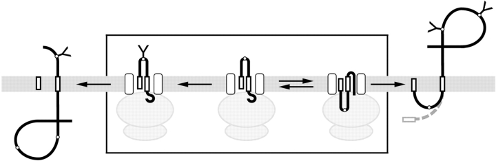
The topology of multispanning membrane proteins in the mammalian endoplasmic reticulum is thought to be dictated primarily by the first hydrophobic sequence. We analyzed the in vivo insertion of a series of chimeric model proteins containing two conflicting signal sequences, i.e., an NH(2)-terminal and an internal signal, each of which normally directs translocation of its COOH-terminal end. When the signals were separated by more than 60 residues, linear insertion with the second signal acting as a stop-transfer sequence was observed. With shorter spacers, an increasing fraction of proteins inserted with a translocated COOH terminus as dictated by the second signal. Whether this resulted from membrane targeting via the second signal was tested by measuring the targeting efficiency of NH(2)-terminal signals followed by polypeptides of different lengths. The results show that targeting is mediated predominantly by the first signal in a protein. Most importantly, we discovered that glycosylation within the spacer sequence affects protein orientation. This indicates that the nascent polypeptide can reorient within the translocation machinery, a process that is blocked by glycosylation. Thus, topogenesis of membrane proteins is a dynamic process in which topogenic information of closely spaced signal and transmembrane sequences is integrated.


Figures

Figure 1
Figure 1
Chimeric proteins and their potential insertion patterns. (A) Schematic representation of the fusion proteins consisting of an NH2-terminal signal sequence (open boxes), a spacer sequence of 20–100 residues, the internal signal-anchor (filled boxes) and the entire COOH-terminal domain of the ASGP receptor H1 with two potential N-glycosylation sites (open circles). Sequences derived from hemagglutinin and the ASGP receptor are shown as open and filled lines, respectively. (B) Possible topologies of the chimeras in the ER membrane. The linear insertion model predicts a cleaved NH2-terminal signal, a translocated spacer, and a cytosolic, unglycosylated COOH terminus (left). Alternatively, the COOH terminus is translocated and glycosylated, the spacer remains cytosolic, and the NH2-terminal signal is uncleaved and either integrated or cytosolic (right). cyt, cytoplasmic side; exo, exoplasmic side. (C) The sequences of the different NH2-terminal signals used. H, hemagglutinin; P, preprolactin; V, prepro-vasopressin-neurophysin II; A and ΔA, ASGP receptor H1 with its complete and a truncated hydrophilic, cytoplasmic domain, respectively. Signal cleavage sites are indicated by a dot.
Figure 2
Figure 2
ER insertion of chimeric proteins containing the NH2-terminal signal of hemagglutinin and the internal signal of the ASGP receptor H1. (A) Transfected COS cells were labeled with [35S]methionine for 30 min. The chimeric proteins were immunoprecipitated and analyzed by SDS-gel electrophoresis and fluorography after deglycosylation with endo H (+) or without treatment (−). (B) Membrane integration tested by alkaline extraction. Transfected COS cells were metabolically labeled and scraped. One half was subjected to alkaline extraction. The resulting supernatant (S) and pellet fractions (P), as well as the other half (the total, T) were then subjected to immunoprecipitation and analyzed by gel electrophoresis and fluorography. As a soluble control, H20−, which lacks a second, internal signal and is secreted into the ER lumen, was analyzed. (C) Translocation efficiency of the hemagglutinin signal in the absence of a second signal sequence. From the constructs H#A, the segment encoding the internal signal of the ASGP receptor H1 was deleted. The resulting H#− constructs were expressed in COS cells, metabolically labeled, and analyzed by immunoprecipitation, gel electrophoresis, and fluorography. The position of molecular weight markers of 35 and 40 kD are indicated.
Figure 3
Figure 3
Insertion of chimeric proteins with different NH2-terminal signals. Chimeric proteins P#A, V#A, A#A, and ΔA#A, which contain the NH2-terminal signals of preprolactin, prepro-vasopressin-neurophysin II, and the full-size or truncated ASGP receptor H1, respectively, were expressed in COS cells and analyzed as described in Fig. 2 A. Asterisks indicate the uncleaved population of P20A. The position of molecular weight markers of 35 and 40 kD are indicated.
Figure 4
Figure 4
Quantitation of transmembrane topologies. Multiple experiments like those shown in Fig. 2 A and Fig. 3 were quantified using a PhosphorImager. The fraction of polypeptides with a glycosylated and thus translocated COOH terminus are plotted versus the length of the spacer separating the two signals. The curves are labeled with the abbreviation for the first signal sequence: H, hemagglutinin (filled circles); P, preprolactin (filled triangles); V, prepro-vasopressin-neurophysin II (filled squares); A, ASGP receptor H1 including the NH2-terminal hydrophilic sequence (open squares); ΔA, ASGP receptor H1 with a truncated NH2-terminal domain (open circles). The means and SDs of three to six determinations are shown.
Figure 5
Figure 5
Two mechanisms for topogenic competition. Competition between the signals for SRP in the cytosol (A) or for the preferred orientation in the translocon (B). See text for details.
Figure 6
Figure 6
Kinetics of SRP recruitment and targeting by the vasopressin and hemagglutinin signals. Polypeptides consisting of the signal sequence of hemagglutinin or vasopressin followed by 55, 75, 95, or 115 residues, including a glycosylation site (H55, H75, etc. and V55, V75, etc.; schematically shown in A) were expressed in COS cells, metabolically labeled for 40 min, immunoprecipitated, incubated with (+) or without endo H (−), and analyzed by SDS-gel electrophoresis and fluorography (B and C). The unglycosylated fraction corresponds to polypeptides that had not been targeted to the ER, yet when the ribosome reached the stop codon and disassembled. The position of molecular weight markers of 9, 14, and 20 kD are indicated.
Figure 7
Figure 7
Effect of glycosylation in the spacer sequence on protein topology. (A) The indicated constructs were expressed in COS cells and analyzed with (+) or without (−) endo H digestion. The ratio of proteins with a translocated COOH terminus (twice glycosylated) to proteins with a translocated spacer sequence (unglycosylated or once glycosylated) shifted in favor of the latter upon insertion of functional glycosylation sites into the spacer segment. (B) Quantitation of experiments as shown in A. The means and SDs of three to five determinations are plotted, except for V40(NMT)A, V60(NIT)A, and H40(NIT)A of which single measurements are shown.
Figure 8
Figure 8
Schematic model of how glycosylation affects topogenesis. See text for details.

References

    1. Andrews D.W., Young J.C., Mirels L.F., Czarnota G.J. The role of the N-region in signal sequence and signal-anchor function. J. Biol. Chem. 1992;267:7761–7769. - PubMed
    1. Beltzer J.P., Fiedler K., Fuhrer C., Geffen I., Handschin C., Wessels H.P., Spiess M. Charged residues are major determinants of the transmembrane orientation of a signal-anchor sequence. J. Biol. Chem. 1991;266:973–978. - PubMed
    1. Blobel G. Intracellular protein topogenesis. Proc. Natl. Acad. Sci. USA. 1980;77:1496–1500. - PMC - PubMed
    1. Brodsky J.L., Goeckeler J., Schekman R. BiP and Sec63p are required for both co- and posttranslational protein translocation into the yeast endoplasmic reticulum. Proc. Natl. Acad. Sci. USA. 1995;92:9643–9646. - PMC - PubMed
    1. Coleman J., Inukai M., Inouye M. Dual functions of the signal peptide in protein transfer across the membrane. Cell. 1985;43:351–360. - PubMed

Publication types

Substances