. 1999 Oct 18;147(2):335-50.
doi: 10.1083/jcb.147.2.335.

Novel roles for saccharomyces cerevisiae mitotic spindle motors

Affiliations

Novel roles for saccharomyces cerevisiae mitotic spindle motors

F R Cottingham et al. J Cell Biol. .

Abstract

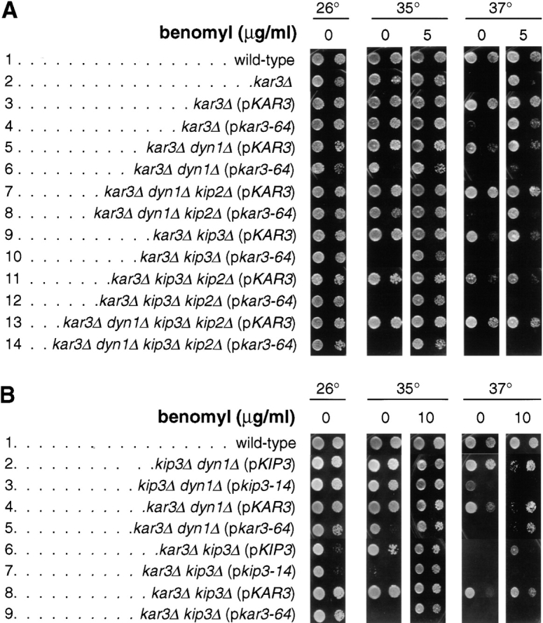
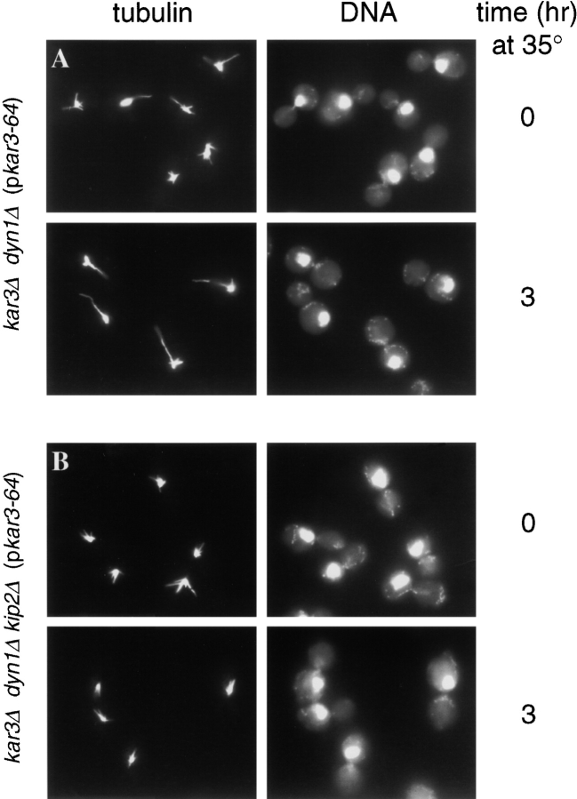
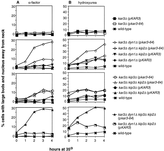
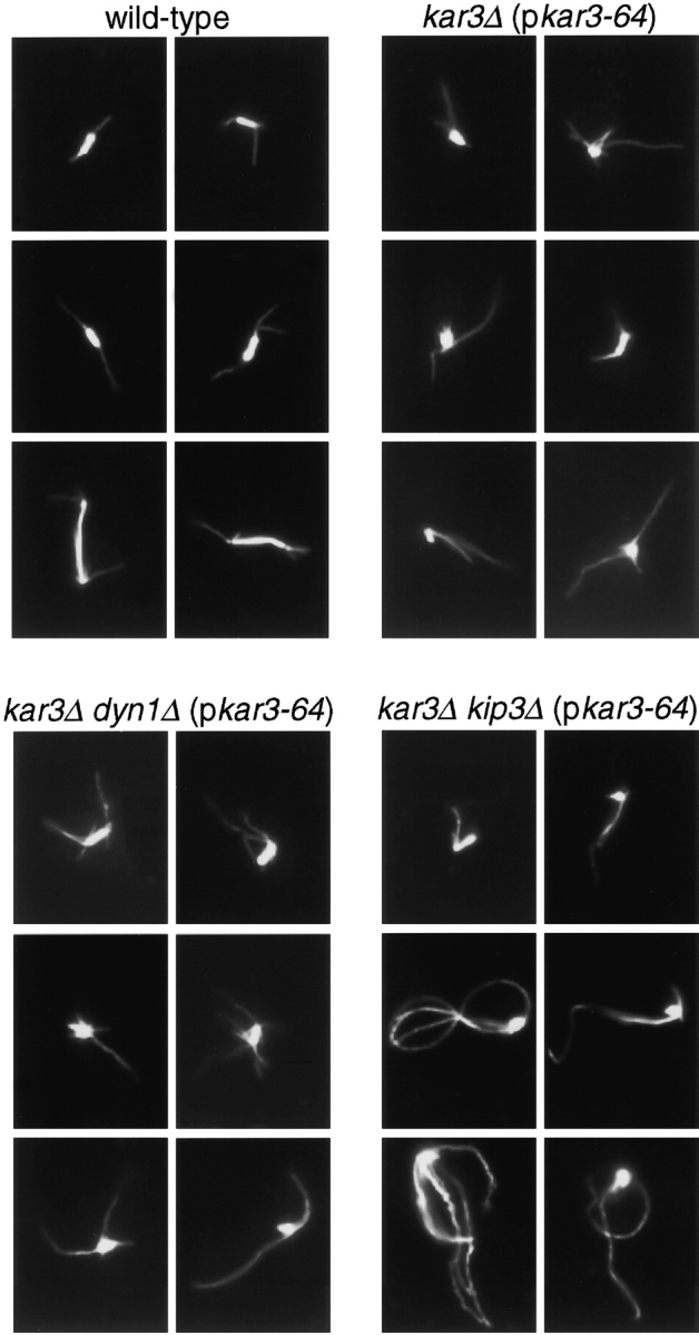
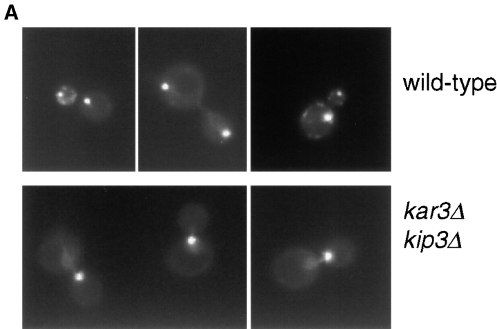
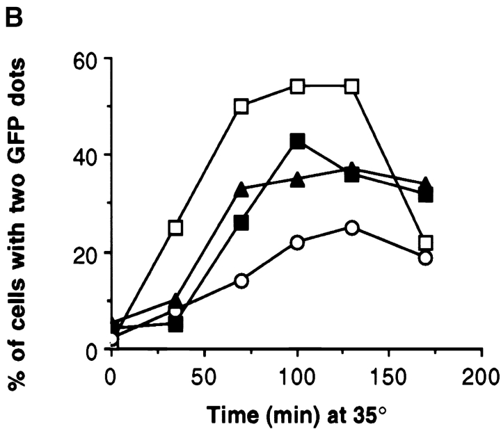
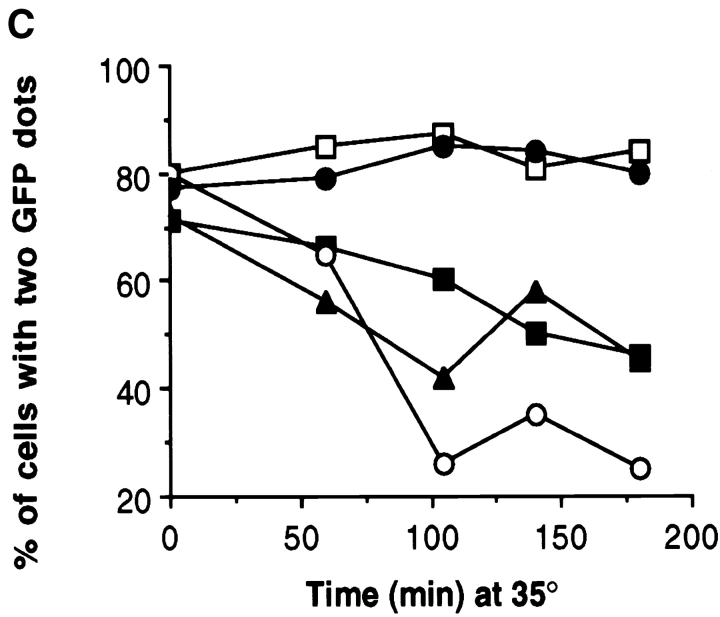
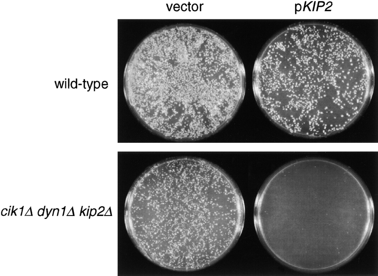
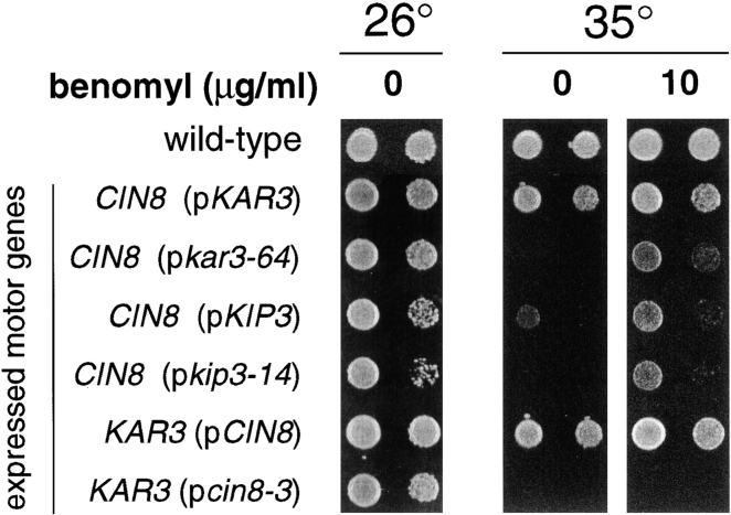
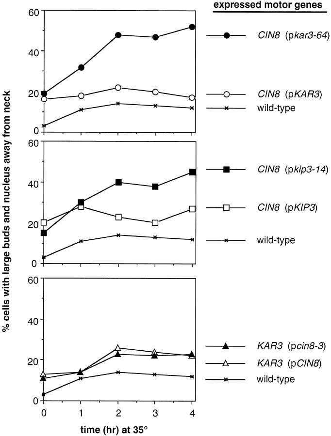
The single cytoplasmic dynein and five of the six kinesin-related proteins encoded by Saccharomyces cerevisiae participate in mitotic spindle function. Some of the motors operate within the nucleus to assemble and elongate the bipolar spindle. Others operate on the cytoplasmic microtubules to effect spindle and nuclear positioning within the cell. This study reveals that kinesin-related Kar3p and Kip3p are unique in that they perform roles both inside and outside the nucleus. Kar3p, like Kip3p, was found to be required for spindle positioning in the absence of dynein. The spindle positioning role of Kar3p is performed in concert with the Cik1p accessory factor, but not the homologous Vik1p. Kar3p and Kip3p were also found to overlap for a function essential for the structural integrity of the bipolar spindle. The cytoplasmic and nuclear roles of both these motors could be partially substituted for by the microtubule-destabilizing agent benomyl, suggesting that these motors perform an essential microtubule-destabilizing function. In addition, we found that yeast cell viability could be supported by as few as two microtubule-based motors: the BimC-type kinesin Cin8p, required for spindle structure, paired with either Kar3p or Kip3p, required for both spindle structure and positioning.


Figures

Figure 1
Figure 1
Growth properties of motor mutant strains. Yeast cells of the indicated genotype were spotted onto solid rich media containing the indicated concentration of benomyl, and incubated at the indicated temperature for 3 d. Two spots appear for each sample: the spot to the right is a 40-fold dilution of the sample on the left. Strains used (p indicates a centromere plasmid-carried gene): (A) wild-type (MAY4360), kar3Δ (MAY5742), kar3Δ (pKAR3) (MAY5022), kar3Δ (pkar3-64) (MAY5398), kar3Δ dyn1Δ (pKAR3) (MAY5218), kar3Δ dyn1Δ (pkar3-64) (MAY5400), kar3Δ dyn1Δ kip2Δ (pKAR3) (MAY5464), kar3Δ dyn1Δ kip2Δ (pkar3-64) (MAY5465), kar3Δ kip3Δ (pKAR3) (MAY5001), kar3Δ kip3Δ (pkar3-64) (MAY5399), kar3Δ kip3Δ kip2Δ (pKAR3) (MAY5269), kar3Δ kip3Δ kip2Δ (pkar3-64) (MAY5522), kar3Δ dyn1Δ kip3Δ kip2Δ (pKAR3) (MAY5774), and kar3Δ dyn1Δ kip3Δ kip2Δ (pkar3-64) (MAY5775). (B) wild-type (MAY589), kip3Δ dyn1Δ (pKIP3) (MAY4924), kip3Δ dyn1Δ (pkip3-14) (MAY4921), kar3Δ dyn1Δ (pKAR3) (MAY5218), kar3Δ dyn1Δ (pkar3-64) (MAY5400), kar3Δ kip3Δ (pKIP3) (MAY4809), kar3Δ kip3Δ (pkip3-14) (MAY4908), kar3Δ kip3Δ (pKAR3) (MAY5001), and kar3Δ kip3Δ (pkar3-64) (MAY5399).
Figure 2
Figure 2
Spindles and nuclei mislocalize in kar3-ts dyn1Δ cells. The nucleus and spindle were drawn away from the proper position at the mother–bud neck after loss of Kar3p function in the absence of dynein, but not in the absence of dynein and Kip2p. (A) kar3Δ dyn1Δ (pkar3-64) cells (MAY5400) were arrested with hydroxyurea at 26°C and then shifted to 35°C for 3 h in the continued presence of hydroxyurea. Samples taken before (top row) and after (bottom row) the temperature shift were processed for antitubulin immunofluorescence (left column) and DAPI staining of DNA (right column). (B) kar3Δ dyn1Δ kip2Δ (pkar3-64) cells (MAY5465) were treated as in A.
Figure 3
Figure 3
Quantitation of nuclear positioning defects. (A) Nuclear positioning defects following α-factor synchronization at 26°C and release to 35°C. At the time points indicated, samples were removed, stained with DAPI, and the percentage of total cells that were large-budded with the nucleus positioned away from the bud neck was determined (defined as those in which the closest distance between the nucleus and the neck was greater than one half of the diameter of the entire nuclear DNA mass). (B) Nuclear positioning defects after hydroxyurea synchronization at 26°C and a shift to 35°C in the continued presence of hydroxyurea (also see Fig. 2). At the time points indicated, cells were analyzed for nuclear position as in A. Strains used are listed in legend of Fig. 1.
Figure 4
Figure 4
Spindle defects caused by loss of Kar3p function. Cell cultures of the genotypes indicated were synchronized with α-factor at 26°C and then released into medium at 35°C. After 60 min, samples were processed for antitubulin immunofluorescence microscopy. Microtubules of six representative cells are shown for each genotype. Note that the top left example of the kar3Δ kip3Δ (pkar3-64) strain represents a rare bipolar spindle observed for cells of this genotype. Strains used are listed in legend of Fig. 1.
Figure 5
Figure 5
Bipolar spindle assembly and structural integrity requires either Kar3p or Kip3p. (A) Cells of the indicated genotypes also expressed the SPB component Nuf2p tagged with GFP. Cultures of the indicated genotypes were synchronized at 26°C with α-factor and released into media at 35°C. After 60 min, samples of live yeast cells were immediately observed under the microscope. Examples of wild-type (top row) and kar3Δ kip3Δ (pkar3-64) (bottom row) cells. (B) Quantitation of spindle assembly proficiency. Cells released from α-factor at 35°C, as in A, were observed and the percentage of cells exhibiting two clearly separated GFP dots was determined. The decreases observed in the later time point are due to cells that have entered the next cell cycle. (C) Quantitation of ability to maintain spindle structural integrity. Cells of the indicated genotype (and expressing Nuf2p-GFP) were synchronized at 26°C with hydroxyurea and shifted to 35°C for the indicated times, at which point the percentage of cells exhibiting two clearly separated GFP dots was determined. Strains used: wild-type (MAY5776), kar3Δ (pkar3-64) (MAY5777), kar3Δ kip3Δ (pkar3-64) (MAY5778), kar3Δ dyn1Δ (pkar3-64) (MAY5779), and kip3Δ (pkip3-14) (MAY6030).
Figure 5
Figure 5
Bipolar spindle assembly and structural integrity requires either Kar3p or Kip3p. (A) Cells of the indicated genotypes also expressed the SPB component Nuf2p tagged with GFP. Cultures of the indicated genotypes were synchronized at 26°C with α-factor and released into media at 35°C. After 60 min, samples of live yeast cells were immediately observed under the microscope. Examples of wild-type (top row) and kar3Δ kip3Δ (pkar3-64) (bottom row) cells. (B) Quantitation of spindle assembly proficiency. Cells released from α-factor at 35°C, as in A, were observed and the percentage of cells exhibiting two clearly separated GFP dots was determined. The decreases observed in the later time point are due to cells that have entered the next cell cycle. (C) Quantitation of ability to maintain spindle structural integrity. Cells of the indicated genotype (and expressing Nuf2p-GFP) were synchronized at 26°C with hydroxyurea and shifted to 35°C for the indicated times, at which point the percentage of cells exhibiting two clearly separated GFP dots was determined. Strains used: wild-type (MAY5776), kar3Δ (pkar3-64) (MAY5777), kar3Δ kip3Δ (pkar3-64) (MAY5778), kar3Δ dyn1Δ (pkar3-64) (MAY5779), and kip3Δ (pkip3-14) (MAY6030).
Figure 6
Figure 6
Electron microscopic analysis of kar3-64 kip3Δ cells. Wild-type (A) and kar3Δ kip3Δ (pkar3-64) cells (B–F) were arrested with hydroxyurea at 26°C, shifted to 35°C for 3 h, fixed, and sectioned for electron microscopy. A shows a normal preanaphase bipolar spindle. All the mutant spindles (B–F) appeared defective. In B and C, spindles have lost bipolarity. Although the poles are still slightly separated, they are not parallel. In D, E, and F spindles have collapsed with poles in a side by side orientation. All SPBs are marked with large arrowheads. Arrow in F marks a pole whose edge was just caught in this section. Small arrowheads mark cytoplasmic microtubules. n, nucleus. Bar, 0.2 μm. Strains used are listed in legend of Fig. 1.
Figure 7
Figure 7
KIP2 is responsible for the growth defect of cik1Δ dyn1Δ cells. Cultures of wild-type (MAY591) and dyn1Δ cik1Δ kip2Δ (MAY5802) cells were split in two and transformed with either the KIP2-containing plasmid pFC50 or an empty LYS2 vector control (pRS317). Selective media (minus lysine) plates were incubated at 26°C for 3 d.
Figure 8
Figure 8
Benomyl rescues the growth defects of cells expressing only CIN8 and either kar3-64 or kip3-14. Yeast cells were spotted onto solid rich media, with or without 10 μg/ml benomyl, and incubated at the indicated temperature for 3 d. The genotypes indicate the only two microtubule motor genes expressed in these cells (except for wild-type). Strains used (Note: p indicates a centromere plasmid-carried gene): wild-type (MAY1089), CIN8 pKAR3 (MAY5903), CIN8 pkar3-64 (MAY5904), CIN8 pKIP3 (MAY5905), CIN8 pkip3-14 (MAY5906), pCIN8 KAR3 (MAY5940), and pcin8-3 KAR3 (MAY5941).
Figure 9
Figure 9
Nuclear positioning proficiency of cells expressing only two microtubule motor genes. Cultures were arrested with hydroxyurea, shifted to 35°C and analyzed as in Fig. 2 and Fig. 3 B. The genotypes indicate the only two microtubule motor genes expressed in these cells (except for wild-type). Strains used are the same as in Fig. 8.

References

    1. Byers B., Goetsch L. Preparation of yeast cells for thin-section electron microscopy. Methods Enzymol. 1991;194:602–608. - PubMed
    1. Carminati J.L., Stearns T. Microtubules orient the mitotic spindle in yeast through dynein-dependent interactions with the cell cortex. J. Cell Biol. 1997;138:629–641. - PMC - PubMed
    1. Cottingham F.R., Hoyt M.A. Mitotic spindle positioning in Saccharomyces cerevisiae is accomplished by antagonistically-acting microtubule motor proteins. J. Cell Biol. 1997;138:1041–1053. - PMC - PubMed
    1. Coue M., Lombillo V.A., McIntosh J.R. Microtubule depolymerization promotes particle and chromosome movement in vitro. J. Cell Biol. 1991;112:1165–1175. - PMC - PubMed
    1. Desai A., Verma S., Mitchison T.J., Walczak C.E. Kin I kinesins are microtubule-destabilizing enzymes. Cell. 1999;96:69–78. - PubMed

Publication types

MeSH terms