. 1999 Oct 18;147(2):447-61.
doi: 10.1083/jcb.147.2.447.

Aggregation of lipid rafts accompanies signaling via the T cell antigen receptor

Affiliations

Aggregation of lipid rafts accompanies signaling via the T cell antigen receptor

P W Janes et al. J Cell Biol. .

Abstract

The role of lipid rafts in T cell antigen receptor (TCR) signaling was investigated using fluorescence microscopy. Lipid rafts labeled with cholera toxin B subunit (CT-B) and cross-linked into patches displayed characteristics of rafts isolated biochemically, including detergent resistance and colocalization with raft-associated proteins. LCK, LAT, and the TCR all colocalized with lipid patches, although TCR association was sensitive to nonionic detergent. Aggregation of the TCR by anti-CD3 mAb cross-linking also caused coaggregation of raft-associated proteins. However, the protein tyrosine phosphatase CD45 did not colocalize to either CT-B or CD3 patches. Cross-linking of either CD3 or CT-B strongly induced tyrosine phosphorylation and recruitment of a ZAP-70(SH2)(2)-green fluorescent protein (GFP) fusion protein to the lipid patches. Also, CT-B patching induced signaling events analagous to TCR stimulation, with the same dependence on expression of key TCR signaling molecules. Targeting of LCK to rafts was necessary for these events, as a nonraft- associated transmembrane LCK chimera, which did not colocalize with TCR patches, could not reconstitute CT-B-induced signaling. Thus, our results indicate a mechanism whereby TCR engagement promotes aggregation of lipid rafts, which facilitates colocalization of LCK, LAT, and the TCR whilst excluding CD45, thereby triggering protein tyrosine phosphorylation.


Figures

Figure 1
Figure 1
Subcellular localization of LCK and CT-B–rhodamine in Jurkat T cells. A, Cells transiently transfected with LCK-GFP were incubated with CT-B–rhodamine and left untreated (control) or treated by cross-linking with anti–CT-B antibody, as described in Materials and Methods. Single confocal sections were taken of either fixed (F) or live (L) cells using fluorescence in GFP and rhodamine channels. B, Control and anti–CT-B patched cells were fixed and stained with anti-LCK antibody for endogenous protein and visualized by confocal microcopy as in A. Bars, 5 μm.
Figure 2
Figure 2
Localization of lipid raft and nonlipid raft markers with respect to CT-B patches. Top row, Jurkat T cells were incubated with fluorescent GM1-BODIPY and then left untreated (control) or treated with CT-B/anti–CT-B patching. Fluorescence of GM1-BODIPY (fluorescein channel) and CT-B–rhodamine was visualized by confocal microscopy. Other rows, Control cells or cells treated with CT-B–rhodamine and anti–CT-B cross-linking were fixed and stained with antibodies against the cell surface proteins CD59, TfR, CD45, and LAT using FITC-conjugated secondary antibodies. Single confocal sections show fluorescence in fluorescein and rhodamine channels. Bar, 5 μm.
Figure 3
Figure 3
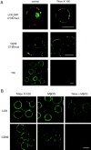
A, Detergent-resistance of proteins associated with CT-B patches. Jurkat cells transfected with LCK-GFP (top row) or left untransfected (other rows) were patched with CT-B–rhodamine plus anti–CT-B. They were then either directly fixed (control) or extracted with 1% Triton X-100 for 5 min on ice before fixation, as indicated. Untransfected cells were stained for CD59 or CD45, using FITC-conjugated secondary antibodies. Fluorescence of GFP and fluorescein (green) or rhodamine (red) was visualized by confocal microscopy, with identical settings for control versus treated samples. For images showing LCK and CD59 localization, red and green images were overlaid to reveal regions of colocalization (appearing yellow) with CT-B. Bars, 10 μm. B, Disruption of detergent-insoluble CT-B–patched proteins by MβCD. Jurkat cells were patched with CT-B/anti–CT-B and then treated with either 1% Triton X-100 for 10 min on ice, 10 mM MβCD for 15 min at 37°C, or MβCD followed by Triton X-100 extraction. The cells were then fixed and stained for LCK or CD59. Confocal images of the various treatments were taken with identical settings to allow comparison of staining. Bar, 10 μm.
Figure 4
Figure 4
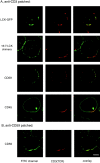
The TCR colocalizes with CT-B patches in Jurkat T cells. A, Cells were treated by patching of CT-B–rhodamine as in earlier figures, and then fixed and stained with anti-CD3 mAb and FITC-conjugated secondary antibody. Bars, 5 μm. B, Cells were CT-B patched, and then fixed directly (control), extracted with 1% Triton X-100 for 5 min on ice, or incubated for 30 min on ice with antibodies against CD3 or TfR to cross-link the receptors before Triton extraction, as indicated. After fixation, the cells were stained for CD3 (top) or TfR (bottom), and confocal images were taken with identical settings to allow comparison of staining before and after treatment. Bar, 10 μm.
Figure 5
Figure 5
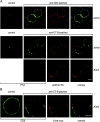
TCR patches resemble lipid raft patches in protein content. A, Cells expressing LCK-GFP (top row), 16:7:LCK (second row), or untransfected Jurkat cells (other rows) were incubated with anti-CD3 mAb followed by anti-Ig Texas red to induce receptor patching. Cells were then fixed and stained as indicated with anti-LCK antiserum followed by FITC-conjugated secondary antibody, or with FITC-conjugated mAbs against CD59 or CD45, to avoid cross-reactivity with anti-CD3 mAb. B, Jurkat cells were incubated with FITC-conjugated anti-CD59 mAb followed by anti-Ig to induce receptor patching. The patched cells were then stained with biotin-conjugated anti-CD3 mAb followed by streptavidin Texas red. Bar, 5 μm.
Figure 6
Figure 6
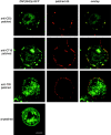
A, TCR and CT-B patching induces tyrosine phosphorylation. Jurkat cells were treated with anti-CD3 mAb and anti-Ig Texas red (top row) or with CT-B–rhodamine plus anti–CT-B (middle row), to induce patching. LCK-deficient JCam-1.6 cells (bottom row) were also patched with CT-B–rhodamine and anti–CT-B. Both patched and control cells were fixed and stained with anti-PTyr polyclonal antibody (top row) or mAb (other rows). Confocal images of CD3-patched or CT-B–patched cells were taken with identical settings to allow comparison of both control versus treated cells and of Jurkat versus JCam-1.6 cells. B, TCR colocalization with CT-B is independent of LCK. Control and CT-B–patched JCam-1.6 cells were fixed and stained with anti-CD3 antibody and visualized by confocal microscopy. Bars, 5 μm.
Figure 7
Figure 7
Cytoplasmic ZAP-70(SH2)2-GFP is relocalized to TCR- and CT-B–cross-linked membrane patches. Jurkat cells were transiently transfected with ZAP-70(SH2)2-GFP and incubated overnight to allow expression. They were then patched with anti-CD3 mAb plus anti-Ig Texas red, CT-B–rhodamine plus anti–CT-B, or anti-TfR mAb plus anti-Ig Texas red, as indicated. After fixation, the cells were analyzed by confocal microscopy. Bar, 5 μm.
Figure 8
Figure 8
CT-B patching induces early TCR signaling events. A, Jurkat cells were treated with CT-B followed by anti–CT-B antibody to induce patching, or stimulated for 5 min with anti-CD3 antibody. Lysates of control and treated cells were analyzed by Western blotting with anti-PTyr antibody. The 25-kD band, seen only in the anti-CD3 treated lane, probably represents cross-reactivity of the mouse secondary antibody with the stimulating mAb. B, Jurkat cells were treated as in A, or with CT-B alone, and lysed. ZAP-70 was immunoprecipitated from lysates of control or treated cells, and immunoprecipitates were analyzed by Western blotting with anti-PTyr antibody. ZAP-70 content of lysates was also compared by Western blotting with anti-ZAP-70 antibody (bottom). C, Lysates of Jurkat cells, treated as in A, were incubated with GST-Grb2 fusion protein to precipitate tyrosine phosphorylated LAT, and then analyzed by Western blotting with anti-PTyr antibody. LAT content of lysates was also compared by Western blotting with anti-LAT antibody (bottom).
Figure 9
Figure 9
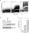
CT-B patching induces signaling pathways downstream from the TCR. A, Ca2+ flux. Jurkat cells were loaded with the Ca2+-binding agent Indo 1, and then treated on ice with CT-B alone (control) or followed by anti–CT-B. Levels of intracellular free Ca2+ were monitored with time by FACS analysis as the cells were warmed to 37°C to induce patching. Indo 1-loaded cells were also stimulated with anti-CD3 mAb as a positive control. B, ERK activation. Jurkat cells were treated with CT-B followed by anti–CT-B, with both agents alone, or stimulated for 5 min with anti-CD3 mAb or PMA. Lysates of control and treated cells were analyzed by Western blotting with antibodies specific for phosphorylated (active) ERK-1 and ERK-2 (top), or for total ERK-2 as a control (bottom). C, NFAT stimulation. Jurkat cells transiently transfected with an NFAT-luciferase reporter construct were treated with CT-B and anti–CT-B to induce lipid raft patching, or stimulated with anti-CD3 antibody. Control and treated cells were incubated overnight before lysis, and lysates were assayed for luciferase activity. The mean of duplicate treatments are shown (± SEM).
Figure 10
Figure 10
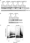
Analysis of CT-B patch-induced signaling in mutant Jurkat cell lines. A, The Jurkat-derived cell lines J.RT3-T3.5 (JRT, TCR/CD3 deficient), JCam-1.6 (JCam, LCK deficient), J45.01 (J45, CD45 deficient), and P116 (ZAP-70 deficient) were compared with parental Jurkat cells for ERK activation after treatment. Cells were treated by CT-B/anti–CT-B patching, with anti-CD3 antibody, or with phorbol ester (PMA) as a positive control. Control and treated cells were assayed for ERK activation as in Fig. 9 B. B, LAT-deficient JCam2 cells (LAT neg) and cells reconstituted with exogenous LAT (LAT pos) were assayed for ERK activation after CT-B or CD3 cross-linking, or PMA stimulation as in A. C, JCam-1.6 cells stably expressing the CD16:CD7:LCK transmembrane chimera (16:7:LCK) and Jurkat cells were loaded with Indo 1 and incubated with CT-B on ice. They were then monitored for intracellular Ca2+ flux, as in Fig. 9 A. Goat anti–CT-B was added to induce CT-B patching of both cell lines at the time shown. For 16:7:LCK cells, mouse anti-CD16 and goat anti–mouse antibodies were then added, followed by rabbit anti–goat (αIg) to cross-link the chimera with CT-B.

References

    1. Anderson R.G.W. The caveolae membrane system. Annu. Rev. Biochem. 1998;67:199–225. - PubMed
    1. Boniface J.J., Rabinowitz J.D., Wulfing C., Hampl J., Reich Z., Altman J.D., Kantor R.M., Beeson C., McConnell H.M., Davis M.M. Initiation of signal transduction through the T cell receptor requires the peptide multivalent engagement of MHC ligands. Immunity. 1998;9:459–466. - PubMed
    1. Brdicka T., Cerny J., Horejsi V. T cell receptor signalling results in rapid tyrosine phosphorylation of the linker protein LAT present in detergent-resistant membrane microdomains. Biochem. Biophys. Res. Commun. 1998;248:356–360. - PubMed
    1. Brown D. The tyrosine kinase connectionhow GPI-anchored proteins activate T cells. Curr. Opin. Immunol. 1993;5:349–354. - PubMed
    1. Brown D.A., Rose J.K. Sorting of GPI-anchored proteins to glycolipid-enriched membrane subdomains during transport to the apical cell surface. Cell. 1992;68:533–544. - PubMed

Publication types