. 1999 Nov;67(11):5676-82.
doi: 10.1128/IAI.67.11.5676-5682.1999.

Expression and characterization of the Mycobacterium tuberculosis serine/threonine protein kinase PknB

Affiliations

Expression and characterization of the Mycobacterium tuberculosis serine/threonine protein kinase PknB

Y Av-Gay et al. Infect Immun. 1999 Nov.

Abstract

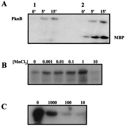
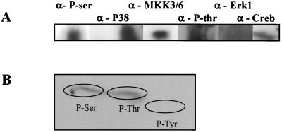
PknB is a member of the newly discovered eukaryotic-like protein serine/threonine kinase (PSTK) family of proteins. The pknB gene was cloned and expressed in Escherichia coli. The active recombinant protein was purified and shown to be reactive with antiphosphoserine antibodies, as well as with antibodies to the phosphorylated eukaryotic Ser/Thr kinases mitogen-activated protein kinase kinase 3 and 6, P38, and Creb. In vitro kinase assays demonstrated that PknB is a functional kinase that is autophosphorylated on serine/threonine residues and is also able to phosphorylate the peptide substrate myelin basic protein. Analysis of pknB expression in Mycobacterium tuberculosis indicates the presence of pknB mRNA in (i) organisms grown in vitro in bacteriological media, (ii) a murine macrophage in vitro infection model, and (iii) in vivo alveolar macrophages from a patient with tuberculosis.


Figures

FIG. 1
FIG. 1
Chromosomal location and cloning of PknB. (A) Schematic map describing the chromosomal location of the pknB open reading frame. (B) PCR amplification of pknB by using gene-specific primers as described in Materials and Methods. Lanes: 1, lambda HindIII molecular size markers; 2, PCR without 5′ primer; 3, PCR without 3′ primer; 4, PCR without template DNA; 5, complete reaction using M. tuberculosis H37Rv DNA as the template. (C) The expression plasmid pYA102.
FIG. 2
FIG. 2
PknB expression. (A) SDS-PAGE analysis of PknB expression in E. coli. Lanes: 1, molecular size markers; 2, negative control pET-22b in E. coli BL21 after IPTG induction; 3, cell extract of pYA102 expressing PknB after IPTG induction; 4, pellet of cell extract of pYA102 expressing PknB after IPTG induction; 5, PknB inclusion bodies after washes with detergent; 6, PknB after urea-DTT treatment; 7, PknB after size-exclusion chromatography. (B) Western analysis using mouse anti-PknB polyclonal antibodies. Lanes: 1, negative control pET-22B; 2, pellet of cell extract of pYA102 expressing PknB after IPTG induction; 3, PknB inclusion bodies; 4, PknB after urea-DTT treatment; 5, PknB after size-exclusion chromatography.
FIG. 3
FIG. 3
In vitro kinase assay. SDS-PAGE analysis of PknB labelled with [γ-32P]ATP. (A) Time course detection of PknB phosphorylation. Lanes: 1, PknB autophosphorylation; 2, MBP phosphorylation mediated by PknB. (B) MnCl2 concentration effect on PknB autophosphorylation. Units are in millimolar concentrations. (C) Effect of serial dilutions of anti-PknB antibodies on MBP phosphorylation by PknB.
FIG. 4
FIG. 4
PknB cross-reactivity with eukaryotic phosphoprotein antibodies and phosphoamino acid analysis. (A) Cross activity of PknB with antibodies against phosphoproteins was determined by SDS–7.5% PAGE analysis followed by Western blot analysis. (B) Phosphoamino acid analysis of autophosphorylated PknB was performed by excision of radioactively labelled PknB from a PVDF membrane followed by acid hydrolysis and two-dimensional thin-layer chromatography. The positions of the unlabeled phosphoamino acids standards are encircled.
FIG. 5
FIG. 5
Transcription analysis of M. tuberculosis pknB. (A) RT-PCR using in vitro-grown M. tuberculosis RNA. Lanes: 1, 1-kb DNA ladder; 2, pknB amplified with cDNA made from M. tuberculosis RNA as a template; 3, pknB amplified from M. tuberculosis genomic DNA preparation; 4, no-DNA-template negative control. (B) Expression upon infection of mouse macrophages. Lanes: 1, 1-kb DNA ladder; 2, cDNA from uninfected control cells after 24 h of incubation; 3, pknB amplified from cDNA from M. tuberculosis infected cells after 24 h of incubation; 4, cDNA from noninfected control cells after 72 h; 5, pknB amplified from cDNA from M. tuberculosis-infected cells after 72 h; 6, pknB amplified from genomic M. tuberculosis DNA; 7, no-DNA template. (C) Expression of pknB in alveolar macrophages from a pulmonary tuberculosis patient. Lanes: 1, 1-kb DNA ladder; 2 to 5, pknB amplified with cDNA derived from alveolar macrophages from a patient suffering from pulmonary tuberculosis; 6, cDNA from noninfected control cells; 7, pknB amplified from genomic M. tuberculosis DNA.

References

    1. Asoh S, Matsuzawa H, Ishino F, Strominger J L, Matsuhashi M, Ohta T. Nucleotide sequence of the pbpA gene and characteristics of the deduced amino acid sequence of penicillin-binding protein 2 of Escherichia coli K12. Eur J Biochem. 1986;160:231–238. - PubMed
    1. Attwood T K, Beck M E, Flower D R, Scordis P, Selley J N. The PRINTS protein fingerprint database in its fifth year. Nucleic Acids Res. 1998;26:304–308. - PMC - PubMed
    1. Av-Gay Y, Davies J. Components of eukaryotic-like protein signaling pathways in Mycobacterium tuberculosis. Microb Comp Genomics. 1997;2:63–73.
    1. Begg K J, Donachie W D. Division planes alternate in spherical cells of Escherichia coli. J Bacteriol. 1998;180:2564–2567. - PMC - PubMed
    1. Brozna J P, Horan M, Rademacher J M, Pabst K M, Pabst M J. Monocyte responses to sulfatide from Mycobacterium tuberculosis: inhibition of priming for enhanced release of superoxide, associated with increased secretion of interleukin-1 and tumor necrosis factor alpha, and altered protein phosphorylation. Infect Immun. 1991;59:2542–2548. - PMC - PubMed

Publication types

MeSH terms

Substances

LinkOut - more resources