

Genomic analysis reveals variation between Mycobacterium tuberculosis H37Rv and the attenuated M. tuberculosis H37Ra strain
- PMID: 10531227
- PMCID: PMC96953
- DOI: 10.1128/IAI.67.11.5768-5774.1999
Genomic analysis reveals variation between Mycobacterium tuberculosis H37Rv and the attenuated M. tuberculosis H37Ra strain
Erratum in
- Infect Immun 2000 Jan;68(1):427
Abstract
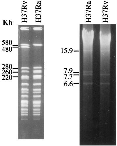
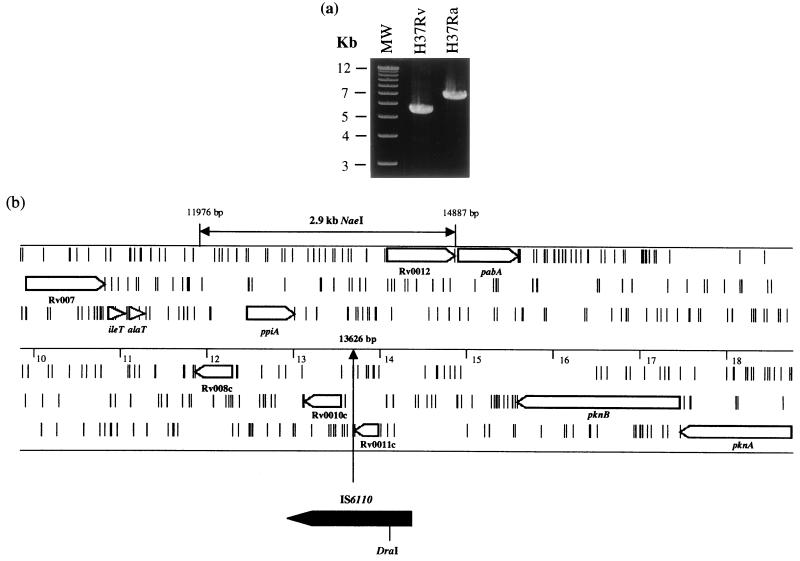
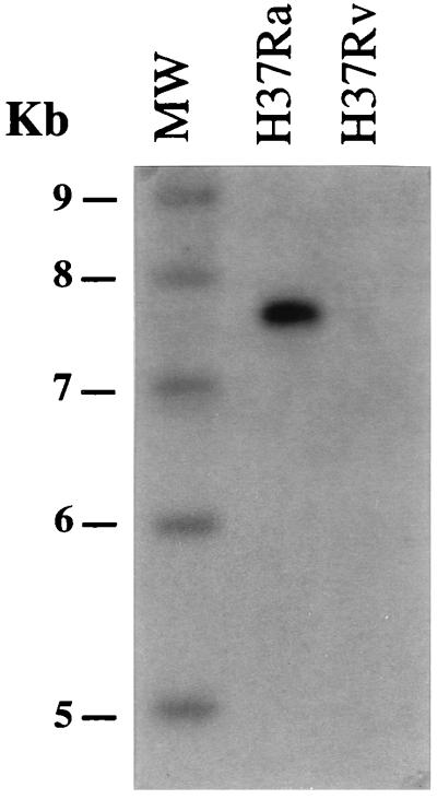
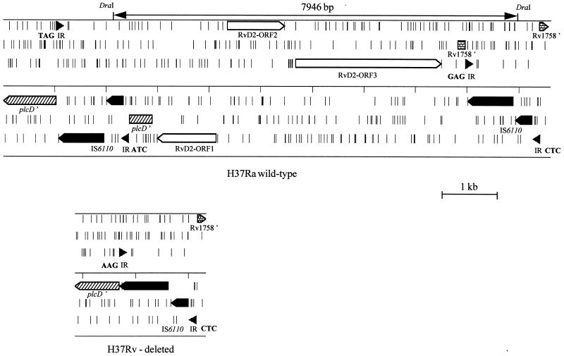
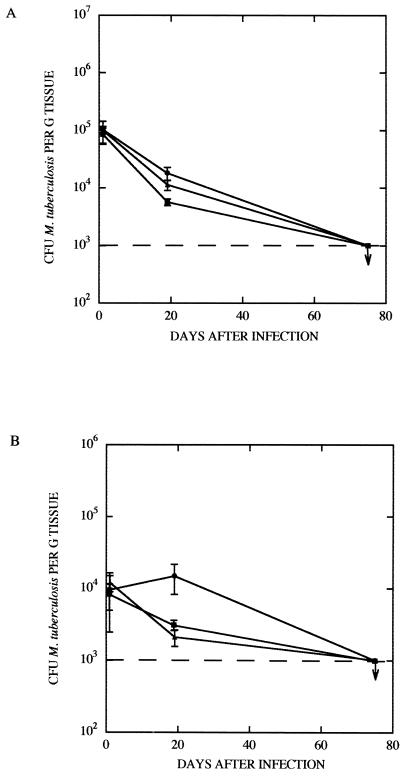
Mycobacterium tuberculosis H37Ra is an attenuated tubercle bacillus closely related to the virulent type strain M. tuberculosis H37Rv. Despite extensive study, the reason for the decreased virulence of M. tuberculosis H37Ra has not been determined. A genomic approach was therefore initiated to identify genetic differences between M. tuberculosis H37Rv and M. tuberculosis H37Ra as a means of pinpointing the attenuating mutation(s). Digestion with the rare-cutting restriction endonuclease DraI revealed two polymorphisms between the strains: a 480-kb fragment in M. tuberculosis H37Rv was replaced by two fragments of 220 and 260 kb in M. tuberculosis H37Ra, while there was a approximately 7.9-kb DraI fragment in M. tuberculosis H37Ra that had no counterpart in M. tuberculosis H37Rv. As the M. tuberculosis insertion sequence IS6110 contains a single DraI restriction site, it was considered possible that these polymorphisms were the result of IS6110 transposition events in M. tuberculosis H37Ra, events that may have inactivated virulence genes. The 7.9-kb polymorphism was found to be due to the presence of the previously described H37Rv RvD2 deletion in M. tuberculosis H37Ra, with sequence analysis suggesting an IS6110-mediated deletion mechanism for loss of RvD2. Three other IS6110-catalyzed deletions from the M. tuberculosis H37Rv chromosome (RvD3 to RvD5) were also identified, suggesting that this mechanism plays an important role in genome plasticity in the tubercle bacilli. Comparative mapping and sequencing revealed that the 480-kb polymorphism was due to an IS6110 insertion in M. tuberculosis H37Ra near oriC. Complementation of M. tuberculosis H37Ra with a 2.9-kb restriction fragment from M. tuberculosis H37Rv that encompassed the IS6110 insertion did not increase the survival of recombinant M. tuberculosis H37Ra in mice. In conclusion, this study describes the presence and mechanisms of genomic variation between M. tuberculosis H37Ra and M. tuberculosis H37Rv, although the role that they play in the attenuation of M. tuberculosis H37Ra is unclear.
Figures
References
-
- Brosch, R., and S. T. Cole. 1999. [Online.] http://www.pasteur.fr/units/Lgmb/. [17 September 1999, last date accessed.]
-
- Calmette A, Guerin C. Sur quelques propriétés du bacille tuberculeux d'origine, cultivé sur la bile de boeuf glycérinée. C R Acad Sci. 1909;149:716–718.
-
- Cole S T, Brosch R, Parkhill J, Garnier T, Churcher C, Harris D, Gordon S V, Eiglmeier K, Gas S, Barry III C E, Tekaia F, Badcock K, Basham D, Brown D, Chillingworth T, Connor R, Davies R, Devlin K, Feltwell T, Gentles S, Hamlin N, Holroyd S, Hornsby T, Jagels K, Barrell B G, et al. Deciphering the biology of Mycobacterium tuberculosis from the complete genome sequence. Nature. 1998;393:537–544. - PubMed
-
- Cole S T, Smith D. Toward mapping and sequencing the genome of Mycobacterium tuberculosis. In: Bloom B R, editor. Tuberculosis: pathogenesis, protection and control. Washington, D.C: American Society for Microbiology; 1994. pp. 227–238.
Publication types
MeSH terms
Substances
Associated data
- Actions
Grants and funding
LinkOut - more resources
Full Text Sources
Other Literature Sources
Research Materials
