. 1999 Nov;67(11):6056-66.
doi: 10.1128/IAI.67.11.6056-6066.1999.

Increased interleukin-1 (IL-1) and imbalance between IL-1 and IL-1 receptor antagonist during acute inflammation in experimental Shigellosis

Affiliations

Increased interleukin-1 (IL-1) and imbalance between IL-1 and IL-1 receptor antagonist during acute inflammation in experimental Shigellosis

J Arondel et al. Infect Immun. 1999 Nov.

Abstract

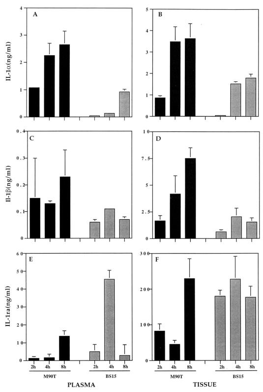
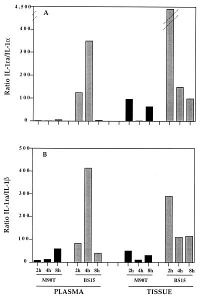
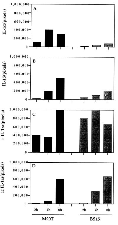
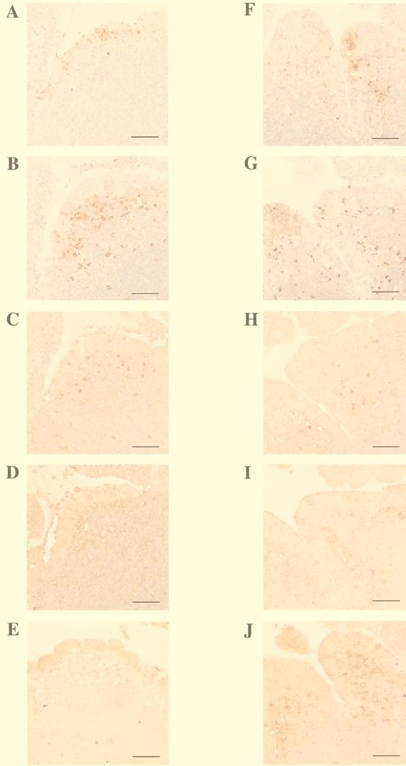
Infection by the enteric bacterial pathogen Shigella results in intense mucosal inflammation and destruction of the colonic and rectal epithelium in infected humans. Initial bacterial translocation occurs through the follicle-associated epithelium. Previous experiments suggest that interleukin-1 (IL-1) is crucial to trigger inflammation, particularly in the follicular zones. During the first 4 hours of infection in a rabbit ligated-loop model of intestinal invasion, there are two salient characteristics: (i) a high concentration of IL-1alpha and IL-1beta, both in infected Peyer's patch tissue and in the corresponding efferent mesenteric blood, and (ii) a very low level of expression of IL-1 receptor antagonist (IL-1ra). These may reflect a combination of regulation of expression and secretion of IL-1alpha, IL-1beta, and IL-1ra by both resident and recruited phagocytes and the induction of mononuclear phagocyte apoptosis by Shigella. This low IL-1ra/IL-1 ratio likely accounts for the rapid, uncontrolled inflammation characteristic of shigellosis.


Figures

FIG. 1
FIG. 1
Shown are IL-1α levels in plasma (A) and in Peyer's patch tissue (B), IL-1β levels in plasma (C) and in Peyer's patch tissue (D), and IL-1ra levels in plasma (E) and in Peyer's patch tissue (F). Cytokine measurements were performed after 2, 4, and 8 h of infection either by invasive S. flexneri M90T (solid bars) or by its noninvasive-adhesive derivative, BS15 (shaded bars). The plasma was from blood samples taken from the mesenteric vein draining the infected loop containing the Peyer's patch. The data represent means plus standard deviations.
FIG. 2
FIG. 2
IL-1ra/IL-1α (A) and IL-1ra/IL-1β (B) ratios in plasma and Peyer's patch tissue during infection with M90T (solid bars) or BS15 (shaded bars).
FIG. 3
FIG. 3
Semiquantitative measurement of IL-1α, IL-1β, sIL-1ra, and icIL-1ra products of RT-PCR carried out on Peyer's patch tissue samples after 2, 4, and 8 h of infection with either M90T or BS15. The results are representative of the data obtained with four to six Peyer's patches for each time point. The data have been normalized according to the amplification of the γ-actin reverse-transcribed products that were standardized at 500,000 pixels.
FIG. 4
FIG. 4
Immunoperoxidase labeling of Peyer's patch tissue sections after 4 h of infection with M90T (A to E) or BS15 (F to J). The following antigens are labeled: LPS (A and F), RAM11 (a macrophage-specific marker) (B and G), IL-1α (C and H), IL-1β (D and I), and IL-1ra (E and J). Bars = 10 μm.
FIG. 5
FIG. 5
TUNEL staining of apoptotic cells in the dome area of a lymphoid follicle infected for 4 h either by the invasive isolate, M90T (A), or by the noninvasive-adhesive isolate, BS15 (B). For better evaluation of the density of fluorescein-labeled apoptotic cells in these samples, only the green TUNEL-positive nuclei have been selected and are shown in panel C (derived from panel A) and in panel D (derived from panel B).
FIG. 6
FIG. 6
Scheme summarizing the possible link between macrophage apoptosis in the dome areas of lymphoid follicles infected by a noninvasive (A) or an invasive (B) Shigella strain and the imbalance between IL-1 and IL-1ra accounting for severe inflammation observed at the early stage of experimental shigellosis. The top cell is an M cell, and the bottom cell is a macrophage in each panel.

References

    1. Adam T, Arpin M, Prévost M C, Gounon P, Sansonetti P J. Cytoskeletal rearrangements and the functional role of T-plastin during entry of Shigella flexneri into HeLa cells. J Cell Biol. 1995;129:367–381. - PMC - PubMed
    1. Andersson J, Björk L, Dinarello C A, Towbin H, Andersson U. Lipopolysaccharide induces human interleukin-1 receptor antagonist and interleukin-1 production in the same cell. Eur J Immunol. 1992;22:2617–2623. - PubMed
    1. Andus T, Daig R, Vogl D. Imbalance of the interleukin-1 system in colonic mucosa: association with intestinal inflammation and interleukin-1 receptor antagonist genotype 2. Gut. 1997;41:651–657. - PMC - PubMed
    1. Arend W P. Interleukin-1 receptor antagonist. Adv Immunol. 1993;54:167–227. - PubMed
    1. Arend W P, Joslin F G, Massoni R J. Effects of immune complexes on production by human monocytes of interleukin 1 or an interleukin 1 inhibitor. J Immunol. 1985;134:3868–3875. - PubMed

Publication types

MeSH terms

LinkOut - more resources