. 1999 Nov 1;19(21):9399-411.
doi: 10.1523/JNEUROSCI.19-21-09399.1999.

Disruption of laminin beta2 chain production causes alterations in morphology and function in the CNS

Affiliations

Disruption of laminin beta2 chain production causes alterations in morphology and function in the CNS

R T Libby et al. J Neurosci. .

Abstract

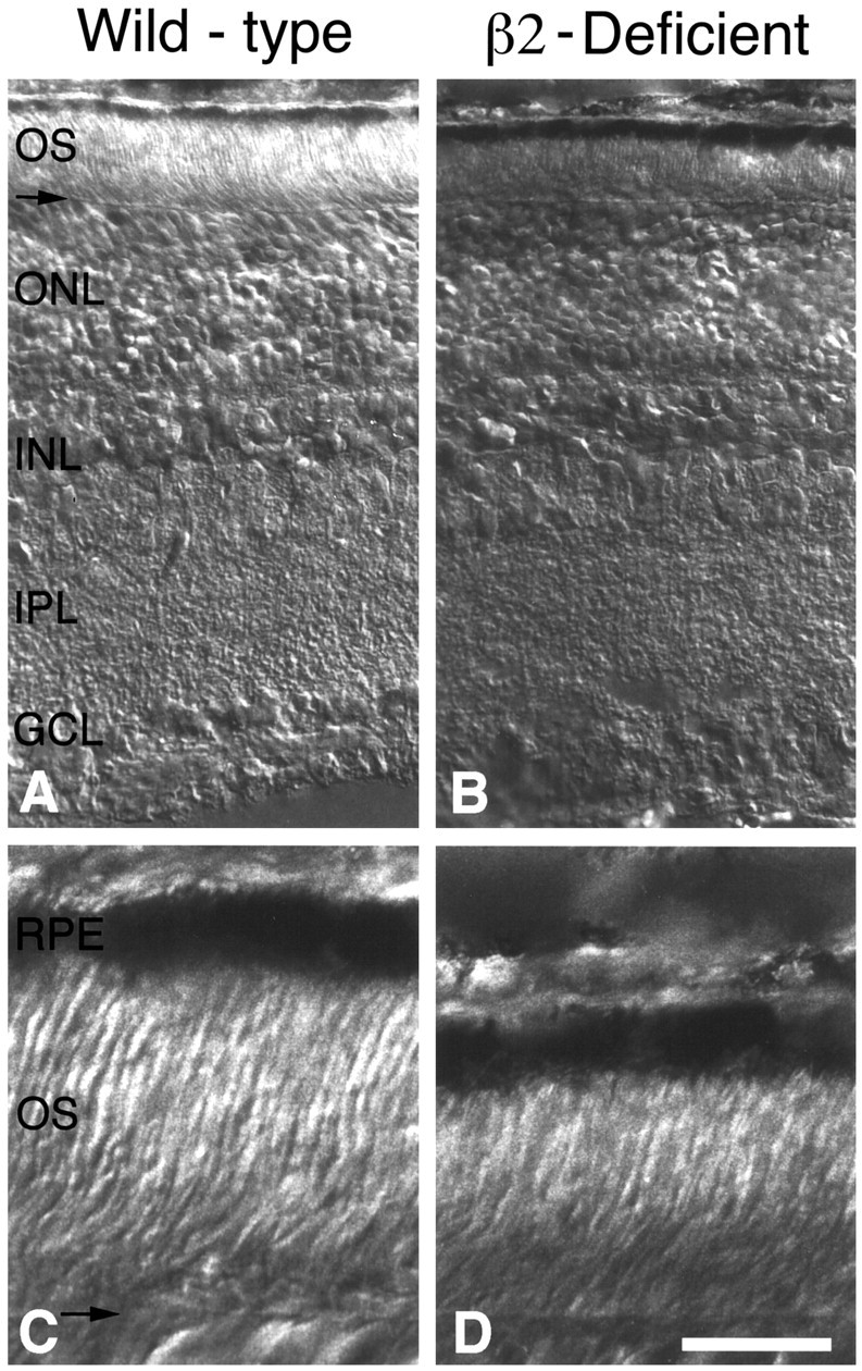
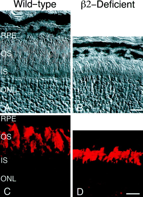
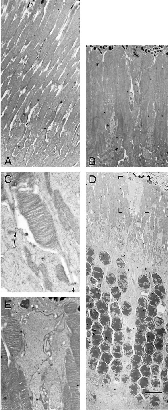
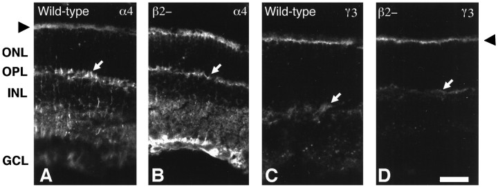
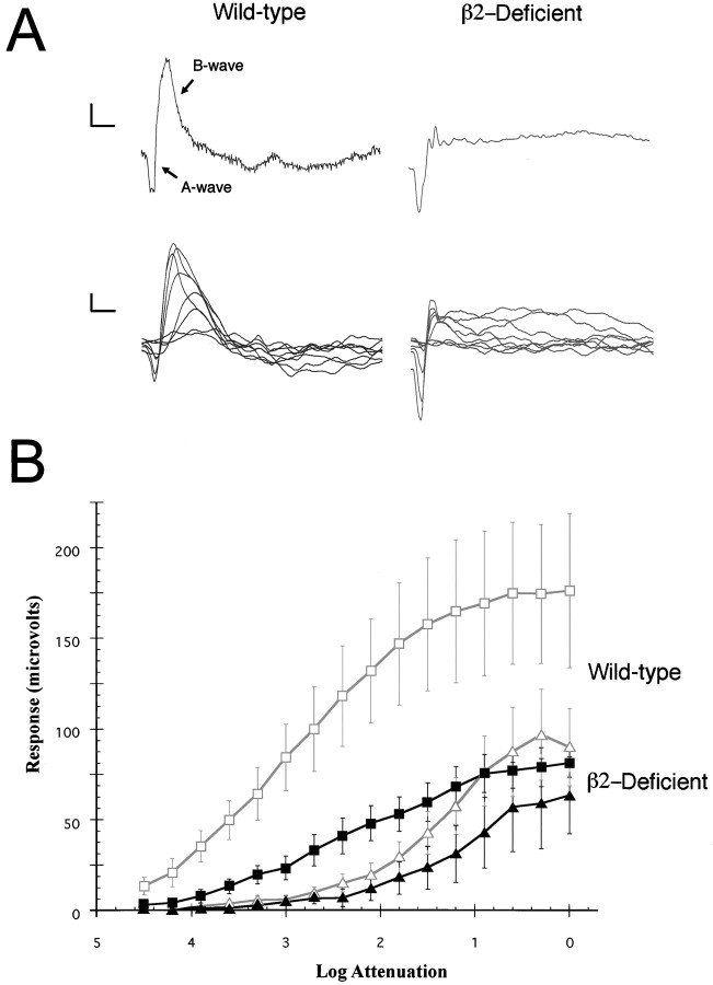
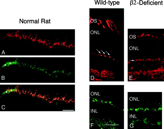
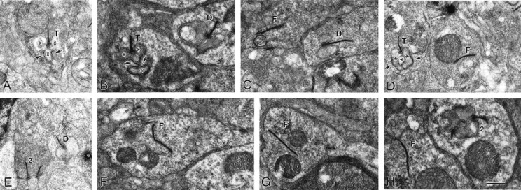
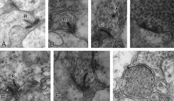
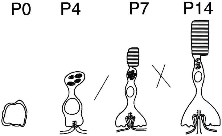
From the elegant studies of Ramon y Cajal (1909) to the current advances in molecular cloning (e.g., Farber and Danciger, 1997), the retina has served as an ideal model for the entire CNS. We have taken advantage of the well described anatomy, physiology, and molecular biology of the retina to begin to examine the role of the laminins, one component of the extracellular matrix, on the processes of neuronal differentiation and synapse formation in the CNS. We have examined the effect of the deletion of one laminin chain, the beta2 chain, on retinal development. The gross development of retinas from laminin beta2 chain-deficient animals appears normal, and photoreceptors are formed. However, these retinas exhibit several pathologies: laminin beta2 chain-deficient mice display abnormal outer segment elongation, abnormal electroretinograms, and abnormal rod photoreceptor synapses. Morphologically, the outer segments are reduced by 50% in length; the outer plexiform layer of mutant animals is disrupted specifically, because only 7% of observed rod invaginating synapses appear normal, whereas the inner plexiform layer is undisturbed; finally, the rate of apoptosis in the mutant photoreceptor layer is twice that of control mice. Physiologically, the electroretinogram is altered; the amplitude of the b-wave and the slope of the b-wave intensity-response function are both decreased, consistent with synaptic disruption in the outer retina. Together, these results emphasize the prominence of the extracellular matrix and, in particular, the laminins in the development and maintenance of synaptic function and morphogenesis in the CNS.


Figures

Fig. 1.
Fig. 1.
Comparison of wild-type and laminin β2 chain-deficient retinas at postnatal day 15. Sections of wild-type (A, C) and laminin β2 chain-deficient littermate (B, D) retinas from areas adjacent to the optic nerve head are shown. There are no gross effects of laminin β2 chain deficiency on the outer nuclear layer (ONL), inner nuclear layer (INL), IPL, or ganglion cell layer (GCL). However, the outer segments (OS) and inner segments of the laminin β2 chain-deficient animal are considerably smaller than those of the wild-type animal, as judged by the distance from the external limiting membrane (arrow) to the retinal pigmented epithelium (RPE). C, D, Enlargements of A and B for clarity. Scale bar: A, B, 50 μm; C, D, 12.5 μm.
Fig. 2.
Fig. 2.
Comparison of wild-type and laminin β2 chain-deficient retinas at postnatal day 25. Sections of wild-type (A, C) and laminin β2 chain-deficient littermate (B, D) retinas, from similar regions, were examined histologically (A, B) or were reacted with an antibody specific for rhodopsin (Ret-P1; C, D). Inner segments (IS) and outer segments (OS) of the wild-type mouse have reached their adult length by postnatal day 25 (A); both inner and outer segments of the laminin β2 chain-deficient mouse are shorter than those of its littermate (B). Wild-type and laminin β2 chain-deficient retinas express rhodopsin and localize it to their outer segments (C, D). However, comparison of rhodopsin expression emphasizes the fact that wild-type OS (C) are significantly longer than those in laminin β2 chain-deficient retinas (D). RPE, Retinal pigmented epithelium; ONL, outer nuclear layer. Scale bars:A, B, 10 μm; C, D, 15 μm.
Fig. 3.
Fig. 3.
Ultrastructural comparison of wild-type and laminin β2 chain-deficient outer retinas at postnatal day 21. Sections of wild-type (A) and laminin β2 chain-deficient (B–E) outer retinas were viewed with transmission electron microscopy. Although photoreceptor inner and outer segments are shorter in the laminin β2 chain-deficient retinas (B), they appear structurally normal, including at the basal bodies (C, arrows). Aberrant processes were occasionally observed in the laminin β2 chain-deficient retinas (D), which extended through the outer nuclear layer and to the retinal pigmented epithelium. The boxed area in Dis shown at higher power in E. Scale bar: A, B, 4.4 μm; C, 1.1 μm; D, 8.5 μm; E, 2.5 μm.
Fig. 4.
Fig. 4.
Other laminin chains are not disrupted by laminin β2 chain deficiency. Unfixed, transverse sections of wild-type and laminin β2 chain-deficient (β2−) littermate retinas were reacted with several antibodies that recognize other laminin chains. The expression pattern of the laminin α4 (A, B) and γ3 (C, D) chains, potential laminin trimer partners of the laminin β2 chain in the neuronal retina, appear to be identical in wild-type (A, C) and laminin β2 chain-deficient (B, D) mice: the laminin α4 and γ3 chains are still present in the subretinal space around inner segments (arrowheads) and in the outer plexiform layer (arrows). The laminin α4 chain (A, B) is also present in both mice throughout the inner retina, through the inner nuclear layer (INL) and the ganglion cell layer (GCL). ONL, Outer nuclear layer. Scale bar, 25 μm.
Fig. 5.
Fig. 5.
Electroretinography of wild-type and laminin β2 chain-deficient retinas. A, Top, The ERGs of a P21, wild-type mouse (left) and a P20, laminin β2 chain-deficient mouse (right) are shown at maximum light intensity. The laminin β2 chain-deficient mouse exhibits a normal a-wave; however, its b-wave is markedly attenuated.Bottom, Intensity–response series (over 4.2 log units, after 21 min of dark adaptation) of a P18, wild-type mouse (left) and a P20, laminin β2 chain-deficient mouse (right). The a- and b-waves of the wild-type mouse increase with increasing levels of light. The laminin β2 chain-deficient mouse shows a similar intensity–response profile, but the b-wave is clearly altered. Calibration: 20 μV (vertical); 100 msec (horizontal). B, The average a-wave amplitudes (triangles) and b-wave amplitudes (squares) for wild-type mice (open symbols) and laminin β2 chain-deficient mice (filled symbols) are shown over a wide range of light intensities. The a-waves of the wild-type mice and the laminin β2 chain-deficient mice are similar; in contrast, the b-waves are attenuated in laminin β2 chain-deficient mice over the entire range.
Fig. 6.
Fig. 6.
The OPL is disrupted in laminin β2 chain-deficient mice. As a mouse-reactive laminin β2 chain antibody was not available, β2 chain expression is shown in rat OPL (A–C). B16 is an antibody that recognizes photoreceptor ribbon synapses (A) and co-localizes with the laminin β2 chain (B) in the OPL (images in A and B are merged inC). Note that the laminin β2 chain is also present in capillaries (*), whereas the B16 antigen is not. D–G, Transverse sections of postnatal day 25 wild-type (D, F) and laminin β2 chain-deficient littermate (E, G) retinas were reacted with a lectin (peanut agglutinin; D, E) that recognizes cone outer segments (OS) and cone photoreceptor terminals (arrows) or an antibody that recognizes all photoreceptor synaptic termini (anti-synaptophysin; F, G) within the OPL, located between the outer nuclear layer (ONL) and the inner nuclear layer (INL). Photoreceptor synapses in the laminin β2 chain-deficient retina appear disorganized. Scale bars, 25 μm.
Fig. 7.
Fig. 7.
Ultrastructural comparison of synapses within the outer plexiform layer of wild-type and laminin β2 chain-deficient retinas at postnatal day 21. Sections of wild-type (A) and laminin β2 chain-deficient (B–H) retinas were viewed with transmission electron microscopy. In the normal retina, rod photoreceptors make synapses, called triads (T), with three postsynaptic elements (*); the ribbon is a presynaptic specialization that marks the active site for release; the three postsynaptic elements invaginate into the base of the photoreceptor such that two horizontal cell dendrites lie laterally, and one bipolar cell dendrite lies centrally. The photoreceptor wraps around the bipolar cell (arrows). In the β2 chain-deficient animal (B–H), several different types of synapses are present. Triads (T) like those in the wild type animal are rare; dyads, with only one or two horizontal cell processes apposed to the ribbon, are more common (D); dyads are seen in wild-type retinas as well (not illustrated). Also common in the β2 chain-deficient retinas are floating synapses (F), wherein a fully formed ribbon, often with vesicles associated, is seen without any postsynaptic element apposed. This type of synapse is seen extremely rarely in the wild-type retina (see Table 1). Finally, occasionally, two ribbons from the same photoreceptor will be apposed to a single postsynaptic element (2); this was also rarely observed in the wild-type animal. Scale bar: A, D, E, 850 nm; B, C, F–H, 630 nm.
Fig. 8.
Fig. 8.
The ultrastructure of the inner plexiform layer of wild-type and β2 chain-deficient animals is similar. Bipolar cell output synapses are ribbon-type synapses (R), whereas amacrine cells make conventional synapses (C) onto other amacrine cells. Two examples of ribbon synapses in wild-type animals (A, E) are shown; apposing the ribbon are one or two postsynaptic processes of unknown origin. Synapses in β2 chain-deficient animals (B–D, F, G) illustrate that the IPL of the mutant animal has both normal ribbon synapses (B, F) and normal conventional synapses (C, D, G), one of which contains a dense-core vesicle (arrowhead). These data suggest that synaptogenesis has proceeded normally in the IPL. Scale bar:A, 340 nm; B–G, 250 nm.
Fig. 9.
Fig. 9.
Disruption of laminin β2 chain production leads to disruption of photoreceptor development. During normal development, photoreceptors begin to develop from an undifferentiated progenitor cell between P0 and P4, at which time they begin to form contacts with processes from interneurons within the outer plexiform layer. The normal developmental program begins to falter in laminin β2 chain-deficient retinas at this stage, with poor development of outer segments and synaptic contacts by P7 and little progress past this point. The net result is that, by P14, the morphogenesis of the photoreceptor is markedly disrupted. This is likely to contribute to the inadequate function that is readily apparent in laminin β2 chain-deficient retinas.

Similar articles

Cited by

References

    1. Altshuler D, Cepko C. A temporally regulated, diffusible activity is required for rod photoreceptor development in vitro. Development. 1992;114:942–957. - PubMed
    1. Arahata K, Ishii H, Hayashi YK. Congenital muscular dystrophies. Curr Opin Neurol. 1995;8:385–390. - PubMed
    1. Balkema GW. A synaptic antigen (B16) is localized in retinal synaptic ribbons. J Comp Neurol. 1991;312:573–583. - PubMed
    1. Balkema GW, Rizkalla R. Ultrastructural localization of a synaptic ribbon protein recognized by antibody B16. J Neurocytol. 1996;25:565–571. - PubMed
    1. Blank M, Koulen P, Blake DJ, Kröger S. Dystrophin and beta-dystroglycan in photoreceptor terminals from normal and mdx3Cv mouse retinae. Eur J Neurosci. 1999;11:2121–2133. - PubMed

Publication types

MeSH terms

LinkOut - more resources