. 1999 Nov 1;19(21):9519-29.
doi: 10.1523/JNEUROSCI.19-21-09519.1999.

Highly specific neuron loss preserves lateral inhibitory circuits in the dentate gyrus of kainate-induced epileptic rats

Affiliations

Highly specific neuron loss preserves lateral inhibitory circuits in the dentate gyrus of kainate-induced epileptic rats

P S Buckmaster et al. J Neurosci. .

Abstract

Patients with temporal lobe epilepsy display neuron loss in the hilus of the dentate gyrus. This has been proposed to be epileptogenic by a variety of different mechanisms. The present study examines the specificity and extent of neuron loss in the dentate gyrus of kainate-treated rats, a model of temporal lobe epilepsy. Kainate-treated rats lose an average of 52% of their GAD-negative hilar neurons (putative mossy cells) and 13% of their GAD-positive cells (GABAergic interneurons) in the dentate gyrus. Interneuron loss is remarkably specific; 83% of the missing GAD-positive neurons are somatostatin-immunoreactive. Of the total neuron loss in the hilus, 97% is attributed to two cell types-mossy cells and somatostatinergic interneurons. The retrograde tracer wheat germ agglutinin (WGA)-apoHRP-gold was used to identify neurons with appropriate axon projections for generating lateral inhibition. Previously, it was shown that lateral inhibition between regions separated by 1 mm persists in the dentate gyrus of kainate-treated rats with hilar neuron loss. Retrogradely labeled GABAergic interneurons are found consistently in sections extending 1 mm septotemporally from the tracer injection site in control and kainate-treated rats. Retrogradely labeled putative mossy cells are found up to 4 mm from the injection site, but kainate-treated rats have fewer than controls, and in several kainate-treated rats virtually all of these cells are missing. These findings support hypotheses of temporal lobe epileptogenesis that involve mossy cell and somatostatinergic neuron loss and suggest that lateral inhibition in the dentate gyrus does not require mossy cells but, instead, may be generated directly by GABAergic interneurons.


Figures

Fig. 1.
Fig. 1.
Nonisotopic in situ hybridization for GAD67-mRNA (A, B) and thionin staining (C, D) in the dentate gyrus of a control rat (A, C) and a kainate-treated rat (B, D). m,Molecular layer; g, granule cell layer;h, hilus; CA3, proximal end of CA3 pyramidal cell layer. Scale bar, 250 μm.
Fig. 2.
Fig. 2.
Septotemporal distribution of GAD67-mRNA-negative hilar neurons (putative mossy cells) in control and kainate-treated (KA-treated) rats. Error bars indicate SEM.
Fig. 3.
Fig. 3.
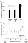
Compared with controls, kainate-treated rats have fewer GAD67-mRNA-positive cells in the hilus (A) and at the temporal pole of the hippocampus (B). Error bars indicate SEM; *p < 0.03,t test.
Fig. 4.
Fig. 4.
Somatostatin-immunoreactivity in the dentate gyrus of the temporal hippocampus in a control rat (A) and a kainate-treated rat (B). Note the lack of somatostatin-immunoreactive interneurons in the hilus (h) of the kainate-treated rat, despite darkly labeled neurons in the CA3 field. g, Granule cell layer;CA3, proximal end of the CA3 pyramidal cell layer. Scale bar, 250 μm.
Fig. 5.
Fig. 5.
There is a significant correlation (r2 = 0.42;F < 0.01, ANOVA) between the number of somatostatin-immunoreactive interneurons and the number of GAD67-mRNA-positive hilar neurons per dentate gyrus in control and kainate-treated (KA-treated) rats.
Fig. 6.
Fig. 6.
WGA-apoHRP-gold injection sites in control (A) and kainate-treated rats (B). Injection sites are indicated by theblack areas. B1, Sections from the kainate-treated rats with the most retrogradely labeled GAD67-mRNA-negative hilar neurons.B2, Sections from the kainate-treated rats with the fewest retrogradely labeled GAD67-mRNA-negative hilar neurons (see Fig. 9). The top section of the left column of A and B2 are from the animals for which the sections are shown in Figure 7, Aand B, respectively. Scale bar, 500 μm.
Fig. 7.
Fig. 7.
Dark-field photomicrographs of silver-intensified WGA-apoHRP-gold-labeled neurons in the dentate gyrus of a control rat (A) and a kainate-treated rat (B). Distance from the injection site (0 mm) is indicated, with negative numbers toward the septal pole and positive numbers toward the temporal pole. The control rat displays numerous retrogradely labeled hilar neurons in sections at least 3.6 mm from the injection site. The kainate-treated rat displays fewer labeled neurons and only in sections near the injection site. Scale bar, 500 μm.— Figure 8. Neurons in the dentate gyrus of control (A) and kainate-treated rats (B) are single- and double-labeled for WGA-apoHRP-gold and GAD67-mRNA. Sections are 1.2 mm from the center of the injection site toward the temporal pole. Arrowheads indicate GAD-positive neurons that were not retrogradely labeled. Arrows indicate retrogradely labeled GAD-negative cells in the hilus (putative mossy cells). Double arrows indicate retrogradely labeled GAD-positive cells. m, Molecular layer;g, granule cell layer; h, hilus. Scale bar, 50 μm.
Fig. 9.
Fig. 9.
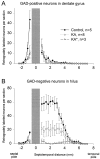
Distribution of retrogradely labeled neurons along the septotemporal axis of the hippocampus. A, Number of retrogradely labeled GAD67-mRNA-positive cells per section in all strata of the dentate gyrus. B, Number of retrogradely labeled GAD67-mRNA-negative hilar neurons (putative mossy cells) per section in control and kainate-treated rats. The injection site, which spans an average of 1.1 mm septotemporally, is centered at 0 septotemporal distance and is indicated by the gray region in the graph. Kainate-treated rats are represented as the entire group (KA) and as a subset of the three animals with the fewest retrogradely labeled GAD-negative hilar neurons (KA*). The control and kainate-treated groups have similar numbers and distributions of retrogradely labeled GAD-positive neurons, which are most abundant in the sections adjacent to the injection site and extend at least 1 mm from the injection site (A). Control rats have numerous GAD-negative hilar neurons in sections extending 4 mm from the injection site (B). KA-treated rats have fewer putative mossy cells than controls, and three KA-treated rats (KA*,triangles, and dashedlines) have almost no retrogradely labeled GAD-negative hilar neurons.

References

    1. Amaral DG. A Golgi study of cell types in the hilar region of the hippocampus in the rat. J Comp Neurol. 1978;182:851–914. - PubMed
    1. Amaral DG, Price JL. An air pressure system for the injection of tracer substances into the brain. J Neurosci Methods. 1983;9:35–43. - PubMed
    1. Amaral DG, Witter MP. The three-dimensional organization of the hippocampal formation: a review of anatomical data. Neuroscience. 1989;31:571–591. - PubMed
    1. Babb TL, Brown WJ, Pretorius J, Davenport C, Lieb J, Crandall PH. Temporal lobe volumetric cell densities in temporal lobe epilepsy. Epilepsia. 1984;25:729–740. - PubMed
    1. Babb TL, Pretorius JK, Kupfer WR, Crandall PH. Glutamate decarboxylase-immunoreactive neurons are preserved in human epileptic hippocampus. J Neurosci. 1989;9:2562–2574. - PMC - PubMed

Publication types

MeSH terms

Substances

LinkOut - more resources