. 1999 Oct 26;96(22):12345-9.
doi: 10.1073/pnas.96.22.12345.

A protein residing at the subunit interface of the bacterial ribosome

Affiliations

A protein residing at the subunit interface of the bacterial ribosome

D E Agafonov et al. Proc Natl Acad Sci U S A. .

Abstract

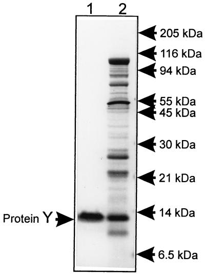
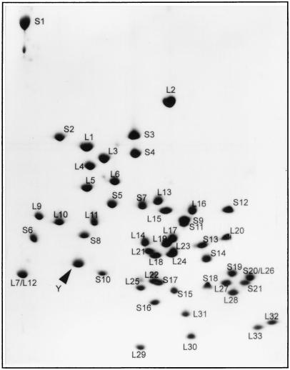
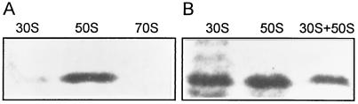
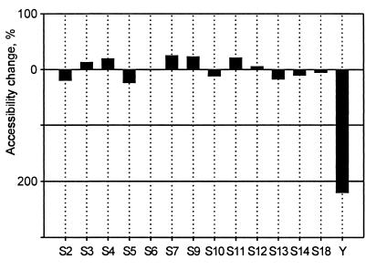
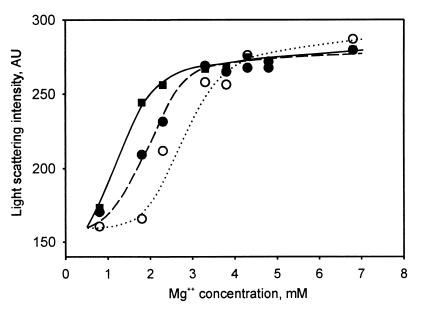
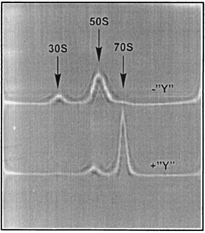
Surface labeling of Escherichia coli ribosomes with the use of the tritium bombardment technique has revealed a minor unidentified ribosome-bound protein (spot Y) that is hidden in the 70S ribosome and becomes highly labeled on dissociation of the 70S ribosome into subunits. In the present work, the N-terminal sequence of the protein Y was determined and its gene was identified as yfia, an ORF located upstream the phe operon of E. coli. This 12.7-kDa protein was isolated and characterized. An affinity of the purified protein Y for the 30S subunit, but not for the 50S ribosomal subunit, was shown. The protein proved to be exposed on the surface of the 30S subunit. The attachment of the 50S subunit resulted in hiding the protein Y, thus suggesting the protein location at the subunit interface in the 70S ribosome. The protein was shown to stabilize ribosomes against dissociation. The possible role of the protein Y as ribosome association factor in translation is discussed.


Figures

Figure 1
Figure 1
SDS/PAGE analysis of purified protein Y. The gel (10% T, 3% C) was stained with Coomassie blue. Lanes: 1, protein Y purified by two-stage column chromatography of the ribosome wash; 2, proteins washed from ribosomes with the dissociation buffer containing 400 mM NaCl.
Figure 2
Figure 2
Separation of individual ribosomal proteins by two-dimensional gel polyacrylamide/urea/SDS electrophoresis. Photograph of Coomassie-stained gel containing 110 μg of total ribosomal protein and ≈5 μg of isolated and purified protein Y (its position is pointed).
Figure 3
Figure 3
Binding of protein Y to ribosomal particles. SDS/PAGE of filtrates passed through the membrane with 100-kDa cutoff. The binding test was performed in buffer D containing 10 mM MgCl2 (A) and 1 mM MgCl2 (B). Type of the ribosomal particle used for binding is indicated above corresponding gel lane. Shown are photographs of Coomassie-stained gels.
Figure 4
Figure 4
Change in protein accessibility as a result of the association of 30S and 50S subunits. The complexes of the 30S subunit with protein Y were labeled with the use of hot tritium bombardment technique before and after the association with the large subunit. Differences in accessibility of individual 30S proteins are shown as percentage of their accessibility in the associated ribosome state (70S), relative to the dissociated ribosome state (30S+50S). Bars plotted below the “zero” line show a decrease in accessibility of a corresponding protein on the 50S subunit attachment.
Figure 5
Figure 5
Mg2+ dependence of ribosome dissociation determined by light scattering at 400 nm. Each sample contained 0.8 μM ribosomes in 150 μl of buffer D. Concentration of MgCl2 in the samples ranged from 0.8 to 6.8 mM. Scattered light intensity is shown in arbitrary units (AU). Samples contained 0.8 μM protein Y (●, dashed line), 2.4 μM protein Y (■, solid line), or no protein Y (○, dotted line).
Figure 6
Figure 6
Sedimentation analysis of protein Y effect on dissociation of the 70S ribosome. Samples contained either 1.25 μM ribosomes without protein Y (the upper pattern, “−Y”) or 1.25 μM ribosomes with 5 μM protein Y (the lower pattern, “+Y”) in 400 μl of buffer D containing 1.8 mM MgCl2. Shown are schlieren optics recording; positions of ribosomal particles are indicated by arrows.

References

    1. Goldanskii V I, Kashirin I A, Shishkov A V, Baratova L A, Grebenshchikov N I. J Mol Biol. 1988;201:567–574. - PubMed
    1. Baratova L A, Grebenshchikov N I, Dobrov E N, Gedrovich A V, Kashirin I A, Shishkov A V, Efimov A V, Jarvekulg L, Radavsky Y L, Saarma M. Virology. 1992;188:175–180. - PubMed
    1. Baratova L A, Grebenshchikov N I, Shishkov A V, Kashirin I A, Radavsky J L, Jarvekulg L, Saarma M. J Gen Virol. 1992;73:229–235. - PubMed
    1. Tsetlin V I, Alyonycheva T N, Shemyakin V V, Neiman L A, Ivanov V T. Eur J Biochem. 1988;178:123–129. - PubMed
    1. Yusupov M M, Spirin A S. FEBS Lett. 1986;197:229–233. - PubMed

Publication types

MeSH terms

LinkOut - more resources