

In Azotobacter vinelandii, the E1 subunit of the pyruvate dehydrogenase complex binds fpr promoter region DNA and ferredoxin I
- PMID: 10535932
- PMCID: PMC22927
- DOI: 10.1073/pnas.96.22.12389
In Azotobacter vinelandii, the E1 subunit of the pyruvate dehydrogenase complex binds fpr promoter region DNA and ferredoxin I
Abstract
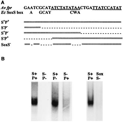
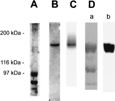
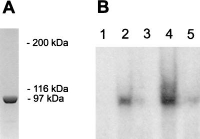
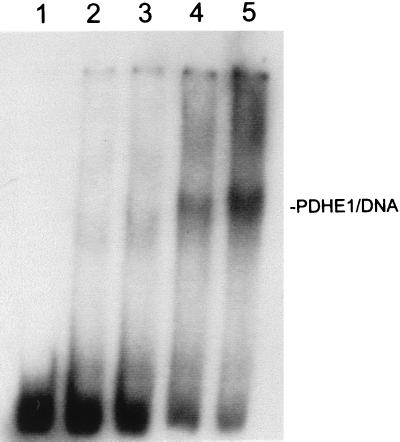
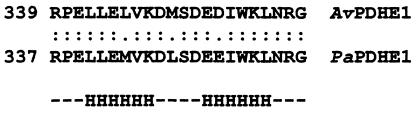
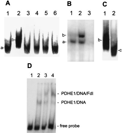
In Azotobacter vinelandii, deletion of the fdxA gene that encodes a well characterized seven-iron ferredoxin (FdI) is known to lead to overexpression of the FdI redox partner, NADPH:ferredoxin reductase (FPR). Previous studies have established that this is an oxidative stress response in which the fpr gene is transcriptionally activated to the same extent in response to either addition of the superoxide propagator paraquat to the cells or to fdxA deletion. In both cases, the activation occurs through a specific DNA sequence located upstream of the fpr gene. Here, we report the identification of the A. vinelandii protein that binds specifically to the paraquat activatable fpr promoter region as the E1 subunit of the pyruvate dehydrogenase complex (PDHE1), a central enzyme in aerobic respiration. Sequence analysis shows that PDHE1, which was not previously suspected to be a DNA-binding protein, has a helix-turn-helix motif. The data presented here further show that FdI binds specifically to the DNA-bound PDHE1.
Figures
Similar articles
-
Identification of a palindromic sequence that is responsible for the up-regulation of NAPDH-ferredoxin reductase in a ferredoxin I deletion strain of Azotobacter vinelandii.J Biol Chem. 1997 May 30;272(22):14454-8. doi: 10.1074/jbc.272.22.14454. J Biol Chem. 1997. PMID: 9162086
-
Complex formation between Azotobacter vinelandii ferredoxin I and its physiological electron donor NADPH-ferredoxin reductase.J Biol Chem. 1999 Jan 29;274(5):2978-87. doi: 10.1074/jbc.274.5.2978. J Biol Chem. 1999. PMID: 9915836
-
E1 component of pyruvate dehydrogenase complex does not regulate the expression of NADPH-ferredoxin reductase in Azotobacter vinelandii.FEMS Microbiol Lett. 2007 Aug;273(2):244-52. doi: 10.1111/j.1574-6968.2007.00797.x. Epub 2007 Jun 16. FEMS Microbiol Lett. 2007. PMID: 17573932
-
Azotobacter vinelandii NADPH:ferredoxin reductase cloning, sequencing, and overexpression.J Biol Chem. 1995 Sep 8;270(36):21258-63. doi: 10.1074/jbc.270.36.21258. J Biol Chem. 1995. PMID: 7673160
-
Atomic structure of the cubic core of the pyruvate dehydrogenase multienzyme complex.Science. 1992 Mar 20;255(5051):1544-50. doi: 10.1126/science.1549782. Science. 1992. PMID: 1549782 Review.
Cited by
-
The Ferredoxin-Like Protein FerR Regulates PrbP Activity in Liberibacter asiaticus.Appl Environ Microbiol. 2019 Feb 6;85(4):e02605-18. doi: 10.1128/AEM.02605-18. Print 2019 Feb 15. Appl Environ Microbiol. 2019. PMID: 30552192 Free PMC article.
-
Functions of the mismatch repair gene mutS from Acinetobacter sp. strain ADP1.J Bacteriol. 2001 Dec;183(23):6822-31. doi: 10.1128/JB.183.23.6822-6831.2001. J Bacteriol. 2001. PMID: 11698371 Free PMC article.
-
The fdxA ferredoxin gene can down-regulate frxA nitroreductase gene expression and is essential in many strains of Helicobacter pylori.J Bacteriol. 2003 May;185(9):2927-35. doi: 10.1128/JB.185.9.2927-2935.2003. J Bacteriol. 2003. PMID: 12700272 Free PMC article.
-
Activation of the Pseudomonas aeruginosa type III secretion system requires an intact pyruvate dehydrogenase aceAB operon.Infect Immun. 2002 Jul;70(7):3973-7. doi: 10.1128/IAI.70.7.3973-3977.2002. Infect Immun. 2002. PMID: 12065547 Free PMC article.
-
The E1beta and E2 subunits of the Bacillus subtilis pyruvate dehydrogenase complex are involved in regulation of sporulation.J Bacteriol. 2002 May;184(10):2780-8. doi: 10.1128/JB.184.10.2780-2788.2002. J Bacteriol. 2002. PMID: 11976308 Free PMC article.
References
-
- Beinert H, Holm R H, Münck E. Science. 1997;277:653–659. - PubMed
-
- GaoSheridan H S, Pershad H R, Armstrong F A, Burgess B K. J Biol Chem. 1998;273:5514–5519. - PubMed
-
- Morgan T V, Lundell D J, Burgess B K. J Biol Chem. 1988;263:1370–1375. - PubMed
-
- Isas J M, Burgess B K. J Biol Chem. 1994;269:19404–19409. - PubMed
Publication types
MeSH terms
Substances
Grants and funding
LinkOut - more resources
Full Text Sources
