. 1999 Oct 26;96(22):12488-93.
doi: 10.1073/pnas.96.22.12488.

Tropomyosin directly modulates actomyosin mechanical performance at the level of a single actin filament

Affiliations

Tropomyosin directly modulates actomyosin mechanical performance at the level of a single actin filament

P VanBuren et al. Proc Natl Acad Sci U S A. .

Abstract

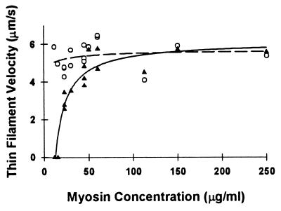
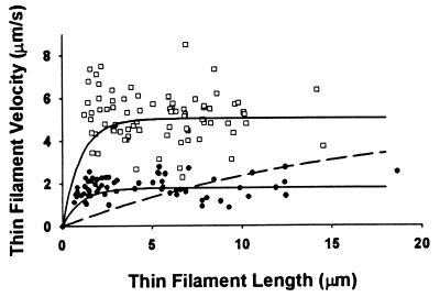
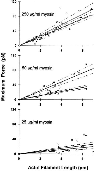
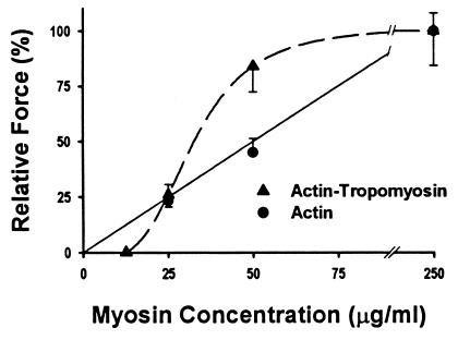
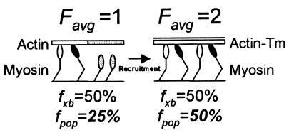
Muscle contraction is the result of myosin cross-bridges (XBs) cyclically interacting with the actin-containing thin filament. This interaction is modulated by the thin filament regulatory proteins, troponin and tropomyosin (Tm). With the use of an in vitro motility assay, the role of Tm in myosin's ability to generate force and motion was assessed. At saturating myosin surface densities, Tm had no effect on thin filament velocity. However, below 50% myosin saturation, a significant reduction in actin-Tm filament velocity was observed, with complete inhibition of movement occurring at 12. 5% of saturating surface densities. Under similar conditions, actin filaments alone demonstrated no reduction in velocity. The effect of Tm on force generation was assessed at the level of a single thin filament. In the absence of Tm, isometric force was a linear function of the density of myosin on the motility surface. At 50% myosin surface saturation, the presence of Tm resulted in a 2-fold enhancement of force relative to actin alone. However, no further potentiation of force was observed with Tm at saturating myosin surface densities. These results indicate that, in the presence of Tm, the strong binding of myosin cooperatively activates the thin filament. The inhibition of velocity at low myosin densities and the potentiation of force at higher myosin densities suggest that Tm can directly modulate the kinetics of a single myosin XB and the recruitment of a population of XBs, respectively. At saturating myosin conditions, Tm does not appear to affect the recruitment or the kinetics of myosin XBs.


Figures

Figure 1
Figure 1
SDS/PAGE and densitometry of a representative pellet and supernatant obtained after a sedimentation assay of an actin–Tm reconstitution. The amount of Tm pelleted with actin implies a 1:6 actin–Tm binding stoichiometry.
Figure 2
Figure 2
Thin filament velocity of actin (○) and actin–Tm (▴) as a function of myosin loading concentration, which in turn determines the motility surface density. Each data point represents an individual experiment in which the mean of at least 20 individual filament velocities are reported. The regression lines for actin–Tm (solid) and actin (dashed) were obtained by fitting the data to the hyperbolic function: y = y0 + (ax)/(b + x); the fit is solely intended to delineate the velocity differences between actin and actin–Tm. The parameters of the fit for actin and actin–Tm were as follows: actin (y0 = 4.71; a = 0.96; b = 19.72); actin–Tm (y0 = −346.91; a = 353.02; b = 0.21).
Figure 3
Figure 3
Velocity of individual actin (□) and actin–Tm filaments (●) as a function of thin filament length and the predicted relationship if the presence of Tm effectively reduces the number of available XBs (dashed curve). The mean velocity of actin and actin–Tm filaments was 5.0 and 1.8 μm/s, respectively, at a myosin concentration of 22.5 μg/ml. At low myosin surface densities, velocity is a function of the number of XBs that can potentially interact with the filament (22). Under these conditions, the determinants of velocity are filament length (i.e., the total number of myosin heads that can interact with the thin filament) and duty cycle (i.e., the fraction of the XB cycle that myosin is strongly bound to actin). The dependence of velocity on filament length is hyperbolic with the asymptote of the relationship achieved, when a sufficient number of XBs interact with the filament to move it at its maximal velocity (17). For illustrative purposes, the solid curves that depict this relationship for actin and actin–Tm are presented and were generated with a defined relationship (17): vmax = avo[1 − (1 − fxb)n], where vmax is the filament velocity, avo is the filament velocity when at least one XB is attached to actin at all times and undergoing its power stroke, fxb is the XB duty cycle, and n is the number of XBs available to interact with the filament. For this model (solid curves), we assumed the avo to be equal to the mean velocities (see above), with fxb = 0.038 and n = 20 heads per micrometer of filament length based on previous studies (17). If Tm effectively reduces the number of available XB heads without having any effect on the kinetics of the XB cycle (i.e., no change in fxb or avo), then one can create a relationship in which the parameters fxb and avo are set at 0.038 and 5.0, respectively, and n is adjusted so that the mean velocity of the fit (over the range of thin filament lengths measured) equals the mean velocity for the actin–Tm data. The dashed curve is this relationship and requires Tm to effectively reduce the number of available XB heads per micrometer of filament length by 92%. If so, then there should have been a profound dependence of velocity on filament length, which was not observed.
Figure 4
Figure 4
Maximal isometric steady-state force per micrometer of thin filament in contact with a myosin-coated surface for actin (▴) and actin–Tm (□) filaments. Experiments were performed with myosin concentrations of 250, 50, and 25 μg/ml. The data were fit by linear regression (solid lines), with 95% confidence limits for the regression indicated by the dashed lines.
Figure 5
Figure 5
Normalized force data for actin ( ● and actin–Tm (▴) as a function of myosin loading concentration, which is related to the surface density. Force for actin–Tm at a myosin concentration of 12.5 μg/ml is extrapolated from the complete inhibition of velocity at this myosin concentration. Saturating myosin surface data were those obtained at a myosin concentration of 250 μg/ml. We have shown (17) that the myosin surface is fully saturated at 100 μg/ml and hence additional myosin loading does not result in additional surface density. In light of this fact, the actin data were fit to a linear regression (solid line) with the surface saturated force measurement being set at a myosin concentration of 100 mg/μl. The actin–Tm data were fit to a sigmoidal regression (dashed line).
Figure 6
Figure 6
Illustration of how a 2× increase in Favg (an ensemble force measurement), resulting from a change in XB recruitment due to the presence of Tm on actin, could be misinterpreted as a change in XB kinetics. In this case, we assume that the total XB population (n) equals 4 and that the XB duty cycle (fxb) is constant at 50% and unchanged in the presence of Tm. In addition, weakly bound XBs are depicted with open heads and strongly bound force-generating XBs are depicted with solid heads and contribute 1 unit of force. With actin alone (Left), it is modeled that the conformation of actin (shaded actin segment) prevents half of the XBs from attaching (shaded detached XBs). Given that only two XBs can interact with actin, and with fxb = 50%, then one XB will generate an Favg =1. However, if one estimates fxb based on Favg and n, then the population-based estimate of fxb is fpop = Favg/n or 25%. With the addition of Tm (Right), all XBs are capable of interacting with actin and Favg = 2. Even though fxb is unchanged, the population-based estimate has increased with fpop = 50%. Without knowing the true XB duty cycle, one could misconstrue the 2× increase in the population-based duty cycle estimate as an effect of Tm on XB kinetics rather than on recruitment. Thus, single molecule studies in the laser trap may be needed to definitively determine the contributions of changes in XB recruitment and kinetics to the observed effects of Tm on actomyosin mechanics.

References

    1. Hill T L, Eisenberg E, Greene L E. Proc Natl Acad Sci USA. 1980;77:3186–3190. - PMC - PubMed
    1. Lehrer S S, Morris E P. J Biol Chem. 1982;257:8073–8080. - PubMed
    1. Lehrer S S. J Muscle Res Cell Motil. 1994;15:232–236. - PubMed
    1. Swartz D R, Moss R L, Greaser M L. Biophys J. 1996;71:1891–1904. - PMC - PubMed
    1. Williams D L J, Greene L E. Biochemistry. 1983;22:2770–2774. - PubMed

Publication types

LinkOut - more resources