. 1999 Oct 26;96(22):12536-41.
doi: 10.1073/pnas.96.22.12536.

A cytomegalovirus-encoded mitochondria-localized inhibitor of apoptosis structurally unrelated to Bcl-2

Affiliations

A cytomegalovirus-encoded mitochondria-localized inhibitor of apoptosis structurally unrelated to Bcl-2

V S Goldmacher et al. Proc Natl Acad Sci U S A. .

Abstract

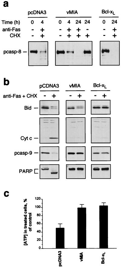
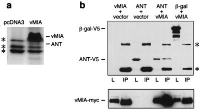
Human cytomegalovirus (CMV), a herpesvirus that causes congenital disease and opportunistic infections in immunocompromised individuals, encodes functions that facilitate efficient viral propagation by altering host cell behavior. Here we show that CMV blocks apoptosis mediated by death receptors and encodes a mitochondria-localized inhibitor of apoptosis, denoted vMIA, capable of suppressing apoptosis induced by diverse stimuli. vMIA, a product of the viral UL37 gene, inhibits Fas-mediated apoptosis at a point downstream of caspase-8 activation and Bid cleavage but upstream of cytochrome c release, while residing in mitochondria and associating with adenine nucleotide translocator. These functional properties resemble those ascribed to Bcl-2; however, the absence of sequence similarity to Bcl-2 or any other known cell death suppressors suggests that vMIA defines a previously undescribed class of anti-apoptotic proteins.


Figures

Figure 1
Figure 1
Identification of a UL37 gene product as an inhibitor of apoptosis. (a) CMV-infected human fibroblasts acquire resistance to Fas- and TNF-α-mediated apoptosis. Cells were infected with CMV(Towne-RIT) (3 plaque-forming units per cell), or left uninfected (N). At the indicated times after infection, cells were exposed for an additional 12 h to medium alone (no treatment), TNF-α + CHX, or anti-Fas antibody + CHX. Viable cells remaining after these treatments were quantified by counting representative fields under a phase-contrast microscope. (b) CMV genomic sequences contained in plasmids with anti-apoptotic activity. (Upper) The structure of the CMV(AD169) genome (5) and UL37 region and associated ORFs are shown schematically. (Lower) CMV DNA inserts in plasmids isolated from three independent library pools (designated 176, 209, and 135). Nucleotide numbering is as described in ref. . Arrows indicate the orientation of inserts relative to the promoter in the expression vector. (c) pUL37x1 suppresses Fas-mediated apoptosis in transfected HeLa cells. Cells were transiently transfected with expression plasmids (1 μg) coding for the following proteins: pUL37x1myc, pUL37x1Δ2–23myc, E1B19K, baculovirus p35, CMV IE1491aa, CMV IE2579aa, or a 1:1 mixture of the plasmids containing IE1 and IE2, or they were transfected with an empty vector (pcDNA3). Twenty-four hours after transfection, the cells were exposed to anti-Fas + CHX for an additional 24 h, and surviving cells were scored under a microscope. (d) (Upper) Structures of pUL37x1, gpUL37M, and gpUL37 produced by alternative splicing. The amino acid numbers used in gpUL37M correspond to those in gpUL37. (Lower) Protection against Fas-mediated apoptosis by pUL37x1myc, gpUL37M, and gpUL37. HeLa cells were transfected with 1 μg of plasmid DNA expressing the indicated proteins and assayed as in c. (e) RNA blot analysis of UL37 exon 3 region transcripts expressed by CMV(AD169) and CMV(Towne-RIT) at 4 and 8 h after infection (Left) or UL37 exon 1 region transcripts expressed by CMV(Towne-RIT) at 4, 8, 24, 48, and 72 h after infection (Right).
Figure 2
Figure 2
HeLa cells constitutively expressing pUL37x1myc are resistant to apoptosis. (a) (Left) Western blot analysis of pUL37x1myc expression in stably transfected HeLa cells with the 9E10 anti-myc antibody. (Right) Expression of cell surface Fas on HeLa clones. Fas levels were examined on cells by flow cytometry, after staining with anti-Fas antibody and a secondary FITC-conjugated antiserum (filled histograms). In control samples, the primary antibody was omitted (open histograms). (b) Resistance of pUL37x1myc HeLa clones to Fas-mediated apoptosis. Cells were exposed to anti-Fas antibody plus CHX, or to CHX only, or to medium alone (untreated), and photographed under a phase-contrast microscope 24 h later. (c) Resistance of pUL37x1myc HeLa clones to apoptosis induced by E1B19K-deficient adenovirus. Cells were infected with Ad2dl250 (3 plaque-forming units per cell), and surviving cells were counted under a microscope at the indicated times. Gray bars, HeLa/pUL37x1myc#3; black bars, HeLa/pcDNA3-A control cells (±SEM, n = 2). (d) Inhibition of doxorubicin-induced apoptosis in pUL37x1myc HeLa cells. Cells were exposed to doxorubicin at the indicated concentrations for 24 h, and the viable cells (as determined by trypan blue exclusion) were counted (±SEM, n = 2; legend as for c).
Figure 3
Figure 3
Mitochondrial localization of pUL37x1 (vMIA). HeLa cells were transiently transfected with plasmids expressing either pUL37x1myc (ac) or pUL37x1Δ2–23myc (d). The cells were permeabilized and stained with either 9E10 anti-myc antibody (a, c, and d, green fluorescence) or anti-mitochondrial antiserum (b, and c, red fluorescence). The yellow coloring (c) resulted from superimposition of red and green fluorescence. Control cells transiently transfected with pcDNA3 did not stain with the 9E10 antibody (not shown). (e and f) Immunoelectron microscopy of ultrathin cryosections of HeLa/pUL37x1myc #3 cells. Cryosections were stained first with 9E10 antibody and then with a secondary antibody-gold conjugate. Two representative fields are shown (M, mitochondria; N, nuclei). HeLa/pcDNA3-A control cells stained with the 9E10 antibody did not reveal specific mitochondrial labeling (not shown). (gi) Time course of vMIA expression in MRC-5 fibroblasts infected with CMV(Towne-RIT). Noninfected cells (g), or cells at 1 day (h) or 2 days (i) after CMV infection were permeabilized and stained with a polyclonal anti-vMIA antibody. No immunofluorescent staining was observed with preimmune serum (not shown).
Figure 4
Figure 4
Effects of vMIA (pUL37x1) expression on biochemical events associated with Fas-mediated apoptosis. (a) HeLa/pcDNA3 cells, HeLa/vMIA cells (pUL37x1myc #3), and HeLa/Bcl-xL cells were treated with CHX, or with anti-Fas antibody plus CHX, or were left untreated. Cell lysates were prepared at the indicated times and procaspase-8 (55 kDa) was detected by Western analysis. (b) Cells were treated with anti-Fas antibody plus CHX for 4 h (+) or were left untreated (−), and the indicated proteins were detected by Western analysis. Cytochrome c was detected in S-100 cell extracts. (c) Cells were treated with anti-Fas antibody plus CHX for 4 h, and the intracellular ATP concentrations were measured by an ATP bioluminescence assay (CLS II, Roche Molecular Biochemicals) and compared with those in control cells (cells exposed to CHX alone). The results are presented as the means ± SEM (n = 4).
Figure 5
Figure 5
Association of vMIA with adenine nucleotide translocator (ANT). (a) Lysates from cells constitutively expressing myc-tagged vMIA, or from control cells (pcDNA3 transfected) were immunoprecipitated with the anti-myc antibody 9E10 covalently linked to Affi-Prep-10 beads. Bound proteins were separated by SDS/PAGE and stained with colloidal Coomassie blue (Novex). The bands designated vMIA and ANT were identified by microsequencing. Asterisks indicate nonspecific bands. (b) (Upper) 293T cells were transiently cotransfected as indicated with expression plasmids (0.5 μg each) coding for vMIAmyc, V5-tagged ANT-1, V5-tagged β-gal, or the empty vector. vMIA was then immunoprecipitated (lanes designated IP) with anti-myc antibody and interacting proteins were detected by Western blot analysis with anti-V5 antibody (Invitrogen). Lanes designated L are samples of the transfected cell lysate taken prior to immunoprecipitation. Asterisks indicate bands of antibody heavy and light chains. (Lower) The blot was reprobed with anti-myc to confirm expression of vMIA-myc.

References

    1. Tschopp J, Thome M, Hofmann K, Meinl E. Curr Opin Genet Dev. 1998;8:82–87. - PubMed
    1. O’Brien V. J Gen Virol. 1998;79:1833–1845. - PubMed
    1. Zhu H, Shen Y, Shenk T. J Virol. 1995;69:7960–7970. - PMC - PubMed
    1. Chee M S, Bankier A T, Beck S, Bohni R, Brown C M, Cerny R, Horsnell T, Hutchison C A, Kouzarides T, Martignetti J A, et al. Curr Top Microbiol Immunol. 1990;154:125–169. - PubMed
    1. Mocarski E S. In: Fields Virology. Fields B N, Knipe D M, editors. Philadelphia: Lippincott-Raven; 1996. pp. 2447–2492.

Substances