. 1999 Oct 26;96(22):12766-71.
doi: 10.1073/pnas.96.22.12766.

Hepatitis C virus and other flaviviridae viruses enter cells via low density lipoprotein receptor

Affiliations

Hepatitis C virus and other flaviviridae viruses enter cells via low density lipoprotein receptor

V Agnello et al. Proc Natl Acad Sci U S A. .

Abstract

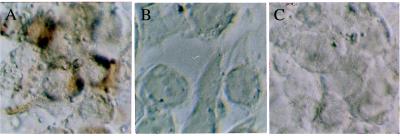
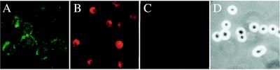
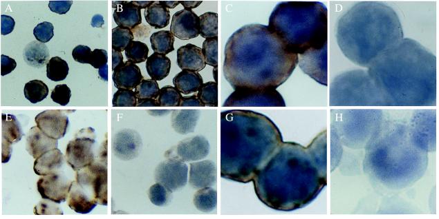
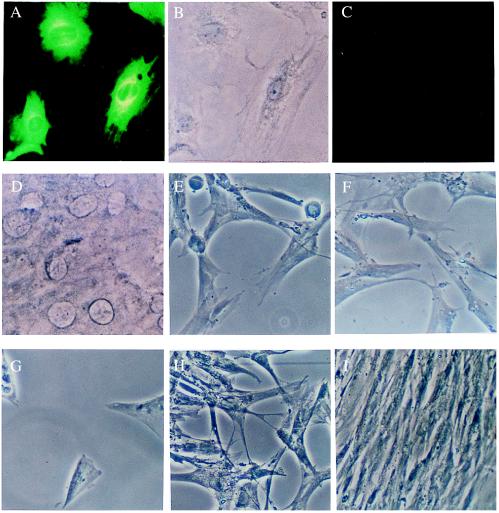
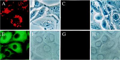
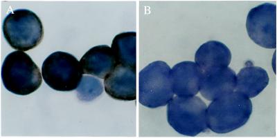
Endocytosis of the Flaviviridae viruses, hepatitis C virus, GB virus C/hepatitis G virus, and bovine viral diarrheal virus (BVDV) was shown to be mediated by low density lipoprotein (LDL) receptors on cultured cells by several lines of evidence: by the demonstration that endocytosis of these virus correlated with LDL receptor activity, by complete inhibition of detectable endocytosis by anti-LDL receptor antibody, by inhibition with anti-apolipoprotein E and -apolipoprotein B antibodies, by chemical methods abrogating lipoprotein/LDL receptor interactions, and by inhibition with the endocytosis inhibitor phenylarsine oxide. Confirmatory evidence was provided by the lack of detectable LDL receptor on cells known to be resistant to BVDV infection. Endocytosis via the LDL receptor was shown to be mediated by complexing of the virus to very low density lipoprotein or LDL but not high density lipoprotein. Studies using LDL receptor-deficient cells or a cytolytic BVDV system indicated that the LDL receptor may be the main but not exclusive means of cell entry of these viruses. Studies on other types of viruses indicated that this mechanism may not be exclusive to Flaviviridae but may be used by viruses that associate with lipoprotein in the blood. These findings provide evidence that the family of LDL receptors may serve as viral receptors.


Figures

Figure 1
Figure 1
Demonstration of the specificity of the ISH method for HCV. (A) HEp 2 cells 24 hours after inoculation with HCV. The brown staining indicates the presence of positive-strand HCV. (B and C) HEp 2 cells incubated with respiratory syncytial virus or adenovirus, respectively, show no staining for HCV using the same ISH method as in A. Note the cytopathic effect of these viruses on the Hep 2 monolayers. Original magnification, ×500.
Figure 2
Figure 2
(A) Demonstration of the LDL receptor on up-regulated G4 cell using anti-LDL receptor antibody (green fluorescent staining). The signal was very weak in ≈30% of G4 cells without up-regulation of the LDL receptor (not shown). (B) Demonstration of the uptake of 5 μg of DiI-LDL by G4 cells that have up-regulated LDL receptors as in A. (C) Inhibition of 5 μg of DiI-LDL endocytosis by 30× excess of unlabeled LDL. (D) Same field as C by phase contrast microscopy. Original magnification, ×500.
Figure 3
Figure 3
(A) HCV ISH of HCV-inoculated G4 cells that were not up-regulated. Dark brown cytoplasmic staining indicates the presence of the virion form of HCV; only ≈30% of the cells are weakly positive. (B) By contrast, HCV was present in most cells in cultures in which the LDL receptor was up-regulated before HCV exposure. Original magnification for A and B, ×500. (C) High-power view of HCV-infected G4 cells with up-regulated LDL receptor as in B. (D) G4 cells prepared as in B and C were pretreated with anti-LDL receptor antibody. Blocking of the receptor by the antibodies decreased the uptake of the virus below the detection limit of ISH. Control antisera (see text) did not have such blocking effect (not shown). Original magnification for C and D, ×1,250. (E) Uptake of HCV by Hep G2 hepatoma cell line is shown by ISH. (F) Blocking the LDL receptor with its specific antibody completely prevented the endocytosis of the virus, as demonstrated by ISH in E. Original magnification for E and F, ×500. (G) Incubation of Daudi cells with HCV-positive serum resulted in positive staining by ISH. (H) Pretreatment of Daudi cells with 2 μM PAO completely inhibited the endocytosis of HCV, as illustrated in G. Original magnification for G and H, ×1250.
Figure 4
Figure 4
(A) Demonstration of infection of BT cell monolayers by cytopathic BVDV (NADL strain) after 72 hours of incubation by immunofluorescence using anti-BVDV (green fluorescent staining). (B) Same field as in A by phase contrast microscopy. (C) Preincubation of BT cell monolayers with anti-LDL receptor antibody has completely prevented the infection (shown in A) of BT cells, as seen after 72 hours of culturing. Same detection method as in A. (D) Phase contrast photomicrograph of the same field as in C. Original magnification for AD, ×500. (E) Inoculation of MRC-5 fibroblasts with HSV resulted in widespread cytolysis and destruction of the monolayer after 48 hours. (F) Pretreatment of the monolayers with anti-LDL receptor antibody, using the same protocol as in C, did not prevent cytolysis and cell death. (G) Incubation of MRC-5 cells with dilutions of VSV also resulted in cytopathy and deterioration of the monolayer. (H) Pretreatment with anti-LDL receptor showed some inhibition of the destruction of the cells. Pretreatment and incubation conditions were as in C and F. (I) Control MRC-5 cell monolayer treated with anti-LDL receptor antibody as in C, F, and H but not inoculated subsequently with virus. Note the uniformity and healthy appearance of these fibroblasts. Original magnification for EI, ×250.
Figure 5
Figure 5
(A) Intense uptake of DiI-LDL by a monolayer of the MDBK cells. (B) Phase contrast microscopy of the same field as A. (C) The lack of endocytosis of DiI-LDL was demonstrated in the CRIB cell line that is resistant to BVDV infection. (D) Same field as in C with phase contrast. (E) Demonstration of the infection of MDBK cells with the NY-1 noncytopathic strain of BVDV after 72 hours of incubation by immunofluorescence using anti-BVDV (green fluorescent staining). (F) Phase contrast picture of E. (G) No BVDV was demonstrated by immunofluorescence in the BVDV-resistant CRIB cells that have been incubated with the virus. Conditions were as in EF. (H) Phase contrast for G. Original magnification for AH, ×500.
Figure 6
Figure 6
(A) Daudi cells inoculated with HGV show the presence of HGV virion in the cytoplasm using ISH specific for this virus. (B) Preincubation of the cells with anti-LDL receptor antibody decreased the uptake of this virus below the detection limit of ISH. Original magnification for A and B, ×1,250.

References

    1. Agnello V, Chung R T, Kaplan L M. N Engl J Med. 1992;327:1490–5. - PubMed
    1. Agnello V. Springer Semin Immunopathol. 1997;19:111–129. - PubMed
    1. Agnello V, Ábel G. Arthritis Rheum. 1997;40:2007–2015. - PubMed
    1. Monti G, Galli M, Invernizzi F, Pioltelli P, Saccardo F, Monteverde A, Pietrogrande M, Renoldi P, Bombardieri S, Bordin G. Q J Med. 1995;88:115–26. - PubMed
    1. Thomssen R, Bonk S, Propfe C, Heermann K H, Kochel H G, Uy A. Med Microbiol Immunol. 1992;181:293–300. - PubMed

Publication types

LinkOut - more resources