. 1999 Oct 26;96(22):12784-9.
doi: 10.1073/pnas.96.22.12784.

Cardioprotection from ischemia by a brief exposure to physiological levels of ethanol: role of epsilon protein kinase C

Affiliations

Cardioprotection from ischemia by a brief exposure to physiological levels of ethanol: role of epsilon protein kinase C

C H Chen et al. Proc Natl Acad Sci U S A. .

Abstract

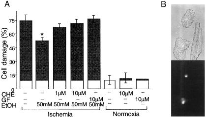
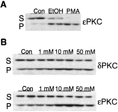
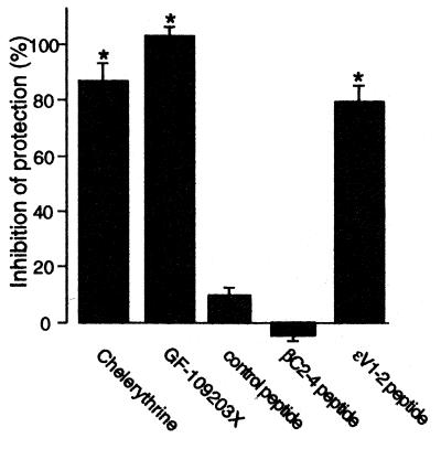
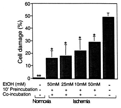
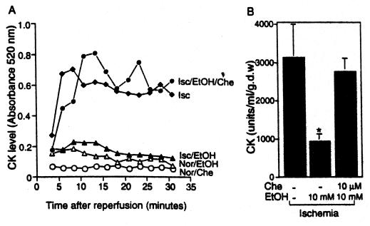
Recent epidemiological studies indicate beneficial effects of moderate ethanol consumption in ischemic heart disease. Most studies, however, focus on the effect of long-term consumption of ethanol. In this study, we determined whether brief exposure to ethanol immediately before ischemia also produces cardioprotection. In addition, because protein kinase C (PKC) has been shown to mediate protection of the heart from ischemia, we determined the role of specific PKC isozymes in ethanol-induced protection. We demonstrated that (i) brief exposure of isolated adult rat cardiac myocytes to 10-50 mM ethanol protected against damage induced by prolonged ischemia; (ii) an isozyme-selective epsilonPKC inhibitor developed in our laboratory inhibited the cardioprotective effect of acute ethanol exposure; (iii) protection of isolated intact adult rat heart also occurred after incubation with 10 mM ethanol 20 min before global ischemia; and (iv) ethanol-induced cardioprotection depended on PKC activation because it was blocked by chelerythrine and GF109203X, two PKC inhibitors. Consumption of 1-2 alcoholic beverages in humans leads to blood alcohol levels of approximately 10 mM. Therefore, our work demonstrates that exposure to physiologically attainable ethanol levels minutes before ischemia provides cardioprotection that is mediated by direct activation of epsilonPKC in the cardiac myocytes. The potential clinical implications of our findings are discussed.


Figures

Figure 1
Figure 1
Ethanol inhibits damage of adult cardiac myocytes induced by in vitro ischemic insult by activating PKC. Adult rat cardiac myocytes were isolated and subjected to control normoxic conditions, simulated ischemia (180 min), or simulated ischemia after a 10-min preincubation with 50 mM ethanol. (A). Membrane fragility to hypotonic solution (85 milliosmolar), a marker of cell damage (34), was determined by uptake of trypan blue. Numbers presented are percentage increase in stained cells attributable to ischemia-induced cell damage as described by the formula formula image Where indicated, chelerythrine (CHE) (1 or 10 μM) or GF109203X (GF) (10 μM) were co-incubated with 50 mM ethanol (EtOH) for 10 min. Cells then were washed twice with buffer and were subjected to simulated ischemia as above. Data are mean ± SEM of seven independent experiments (*, P < 0.05) for the ischemic conditions and three independent experiments for the effect of chelerythrine or GF on membrane fragility. White area represents damage accrued during normoxia. (B) Ischemic injury also was demonstrated by increase in rounded cells and a decrease in rod-shape cells (Upper) and by staining of nuclei with propidium iodide (Lower), previously reported markers of cell damage (38, 39).
Figure 2
Figure 2
Ethanol induces translocation of ɛPKC. Shown is a Western blot analysis of duplicate sets of subcellular fractions from isolated adult rat cardiac myocytes. (A) Myocytes were treated with vehicle (Con), 50 mM ethanol (EtOH), or 100 nM 4β-phorbol 12-myristate 13-acetate (PMA) for 15 min. Soluble and particulate fractions from equal numbers of myocytes (75–100 mg protein per lane) were subjected to SDS/PAGE, were transferred to nitrocellulose, and were probed with an ɛPKC isozyme-selective antibody. Results are representative of two independent experiments. (B) Myocytes were treated with 1 mM, 10 mM, or 50 mM ethanol for 15 min. Soluble and particulate fractions from equal numbers of control (Con) and treated myocytes (75–100 μg protein per lane) were subjected to SDS/PAGE, were transferred to nitrocellulose, and were probed with δPKC or ɛPKC selective antibody. Results are representative of three to four independent experiments.
Figure 3
Figure 3
Inhibition of ethanol protection by an ɛPKC isozyme-selective antagonist peptide, ɛV1-2. Ethanol-induced protection of cardiac myocytes from damage induced by simulated ischemia was determined in the presence of chelerythrine (10 μM), GF109203X (10 μM), the ɛPKC selective inhibitor ɛV1-2, or the cPKC-selective inhibitor βC2-4, each conjugated to the cell-permeable Drosophila Antennapedia carrier peptide and applied at 1 μM. The carrier peptide alone (1 μM) also was used as a negative control. After co-incubation with 50 mM ethanol for 10 min, cells were washed and subjected to simulated ischemia for 180 min. Data, presented as percent inhibition of maximal protection by 50 mM ethanol, are mean ± SEM of three experiments (*, P < 0.05).
Figure 4
Figure 4
Ten millimolar ethanol is sufficient to produce inhibition of ischemic damage. Adult rat cardiac myocytes were isolated and subjected to control normoxic conditions (Normoxia), simulated ischemia (Ischemia), and simulated ischemia after preincubation or preincubation followed by co-incubation with 10–50 mM ethanol as described in Materials and Methods. Cell damage then was determined as in Fig. 1, and data are mean ± SEM of three independent experiments (*, P < 0.05; **, P < 0.005).
Figure 5
Figure 5
Ex vivo exposure of intact heart to 10 mM ethanol is sufficient to produce inhibition of ischemic reperfusion damage. (A) Hearts were perfused without or with 10 mM ethanol (EtOH) for 20 min before the ischemic damage. Where indicated, the PKC inhibitor, chelerythrine (Che) (10 μM) also was perfused. Ischemia-reperfusion injury then was determined by colorimetric reaction of creatine kinase (CK) released from the myocardium as described in Materials and Methods. Numbers presented are OD reading at 520 nm of the 2.5-min fraction collected during the 30-min reperfusion period after a 45-min no-flow ischemia (Isc) or continuous perfusion of oxygenated buffer for the same period of time (Nor). Data are each from a single heart. (B) Ischemia-reperfusion injury is presented as units of CK released during the entire 30-min reperfusion period. Perfusion protocol and treatments of ethanol and chelerythrine was identical as in A. Results are mean ± SEM obtained from each of the three animals (*, P < 0.05).

References

    1. Parratt J R. Trends Pharmacol Sci. 1994;15:19–25. - PubMed
    1. De Labry L O, Glynn R J, Levenson M R, Hermos J A, LoCastro J S, Vokonas P S. J Stud Alcohol. 1992;53:25–32. - PubMed
    1. Ahlawat S K, Siwach S B. Int J Cardiol. 1994;44:157–162. - PubMed
    1. Langer R D, Criqui M H, Reed D M. Circulation. 1992;85:910–915. - PubMed
    1. Kobayashi H, Ashraf M, Rahamathulla P M, Minami M. Pathol Res Pract. 1987;182:810–816. - PubMed

Publication types

MeSH terms

LinkOut - more resources