. 1999 Oct 26;96(22):12804-9.
doi: 10.1073/pnas.96.22.12804.

Autocrine production and action of IL-3 and granulocyte colony-stimulating factor in chronic myeloid leukemia

Affiliations

Autocrine production and action of IL-3 and granulocyte colony-stimulating factor in chronic myeloid leukemia

X Jiang et al. Proc Natl Acad Sci U S A. .

Abstract

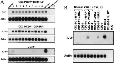
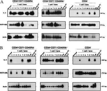
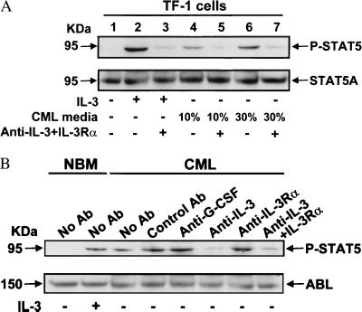
Primitive subsets of leukemic cells isolated by using fluorescence-activated cell sorting from patients with newly diagnosed Ph(+)/BCR-ABL(+) chronic myeloid leukemia display an abnormal ability to proliferate in vitro in the absence of added growth factors. We now show from analyses of growth-factor gene expression, protein production, and antibody inhibition studies that this deregulated growth can be explained, at least in part, by a novel differentiation-controlled autocrine mechanism. This mechanism involves the consistent and selective activation of IL-3 and granulocyte colony-stimulating factor (G-CSF) production and a stimulation of STAT5 phosphorylation in CD34(+) leukemic cells. When these cells differentiate into CD34(-) cells in vivo, IL-3 and G-CSF production declines, and the cells concomitantly lose their capacity for autonomous growth in vitro despite their continued expression of BCR-ABL. Based on previous studies of normal cells, excessive exposure of the most primitive chronic myeloid leukemia cells to IL-3 and G-CSF through an autocrine mechanism could explain their paradoxically decreased self-renewal in vitro and slow accumulation in vivo, in spite of an increased cycling activity and selective expansion of later compartments.


Figures

Figure 1
Figure 1
RT-PCR detection of IL-3 mRNA in CD34+CD71CD45RA, CD34+CD71+CD45RA+, and CD34 cells from 12 CML samples (2,000 cells per subpopulation from each patient). (A) Lane 1 contained PCR products from normal marrow cells, lanes 2–11 from the 10 CML samples with Ph+ LTC-IC, lane 12 from a mouse fibroblast cell line engineered to express human IL-3 (54) (positive control). Lane 13 is the same as lane 12 but with no RT added (negative control), and lane 14 contained water only (another negative control). The same membranes were then washed and rehybridized to a human actin cDNA probe. (B) Lanes 1–3 contained PCR products from normal marrow cells, lanes 4–9 from CML patients 11 and 12 (in which only Ph LTC-IC were detected). Positive and negative controls are the same as in A.
Figure 2
Figure 2
Southern blots showing RT-PCR detection of IL-3, BCR–ABL, and actin transcripts in single CD34+CD71CD45RA, CD34+CD71+CD45RA+, and CD34 cells from one of the CML patients with predominantly Ph+ LTC-IC (no. 10) (A) and one with predominantly Ph LTC-IC (no. 11) (B). The expected sizes, in bp, are indicated for each PCR product. Negative controls are the same as in Fig. 1.
Figure 3
Figure 3
Inhibition of STAT5 phosphorylation in TF-1 cells stimulated by conditioned medium from CML cells (A) and in CD34+ cells from CML patient no. 9 (B), by antibodies to IL-3 and IL-3Rα. In each experiment, a Western blot was performed first with an anti-phospho-STAT5 A/B antibody (P-STAT5, Upper). The membranes were then stripped and reprobed with antibodies to STAT5A (A) and ABL (B) as indicated. Control antibody was the non-blocking anti-IL-3Rα antibody (9F5) (35). For further experimental details, see Methods.
Figure 4
Figure 4
Factor-independent growth of CML cells in culture decreases with their differentiated state. Highly purified CD34+CD71CD45RA (diamonds), CD34+CD71+CD45RA+ (squares), and CD34 (triangle) CML cells (n = 5) or normal marrow cells (circles, n = 3) were cultured at 105 cells per ml in SFM with (solid symbols) or without (open symbols) growth factors (IL-3, IL-6, G-CSF, FL, or SF). Viable cell numbers were determined by hematocytometer counts of trypan blue-excluding cells. Results are expressed as the mean fold-change compared with input values ± SEM. (A) CML patients with predominantly Ph+ LTC-IC (n = 3). (B) CML patients with predominantly Ph LTC-IC (n = 2).
Figure 5
Figure 5
Schematic diagram comparing the predicted effect on different CML cell subpopulations of an autocrine IL-3/G-CSF mechanism based on documented effects of excess exposure to these two growth factors on their phenotypically normal counterparts (37). Dotted arrows indicate the different conditions of growth-factor stimulation compared (supplied exogenously, in the case of the normal cells, or by an autocrine mechanism, in the case of the CML cells). Solid arrows indicate the self-renewal (arched arrows) vs. differentiation (vertical arrows) responses elicited. In each case, the width of the arrow reflects the magnitude of the stimulus or response indicated. The pathology of CML cell expansion in vivo (slow at the level of the leukemic stem-cell compartment and then more rapidly with their progressive differentiation) is consistent with what is observed in vitro when analogous types of very primitive normal hematopoietic cells are exposed to excessive concentrations of IL-3 (±G-CSF).

References

    1. Raskind W H, Fialkow P J. Adv Cancer Res. 1987;49:127–167. - PubMed
    1. Eaves A C, Barnett M J, Ponchio L, Cashman J D, Petzer A, Eaves C J. Stem Cells (Dayton) 1998;16:77–83. - PubMed
    1. Verfaillie C M. In: Stem Cells in Chronic Myelogenous Leukemia. Zon L I, editor. Vol. 4. Philadelphia: W. B. Saunders; 1998. pp. 1079–1114. - PubMed
    1. Eaves C, Cashman J, Eaves A. Leuk Res. 1998;22:1085–1096. - PubMed
    1. Groffen J, Stephenson J R, Heisterkamp N, De Klein A, Bartram C R, Grosveld G. Cell. 1984;36:93–99. - PubMed

Publication types

MeSH terms