. 1999 Oct 26;96(22):12860-5.
doi: 10.1073/pnas.96.22.12860.

Postsynaptic clustering of gamma-aminobutyric acid type A receptors by the gamma3 subunit in vivo

Affiliations

Postsynaptic clustering of gamma-aminobutyric acid type A receptors by the gamma3 subunit in vivo

K Baer et al. Proc Natl Acad Sci U S A. .

Abstract

Synaptic localization of gamma-aminobutyric acid type A (GABA(A)) receptors is a prerequisite for synaptic inhibitory function, but the mechanism by which different receptor subtypes are localized to postsynaptic sites is poorly understood. The gamma2 subunit and the postsynaptic clustering protein gephyrin are required for synaptic localization and function of major GABA(A) receptor subtypes. We now show that transgenic overexpression of the gamma3 subunit in gamma2 subunit-deficient mice restores benzodiazepine binding sites, benzodiazepine-modulated whole cell currents, and postsynaptic miniature currents, suggesting the formation of functional, postsynaptic receptors. Moreover, the gamma3 subunit can substitute for gamma2 in the formation of GABA(A) receptors that are synaptically clustered and colocalized with gephyrin in vivo. These clusters were formed even in brain regions devoid of endogenous gamma3 subunit, indicating that the factors present for clustering of gamma2 subunit-containing receptors are sufficient to cluster gamma3 subunit-containing receptors. The GABA(A) receptor and gephyrin-clustering properties of the ectopic gamma3 subunit were also observed for the endogenous gamma3 subunit, but only in the absence of the gamma2 subunit, suggesting that the gamma3 subunit is at a competitive disadvantage with the gamma2 subunit for clustering of postsynaptic GABA(A) receptors in wild-type mice.


Figures
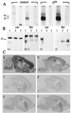
Figure 1
Figure 1
Expression of γ3 subunit and reconstitution of BZ binding sites in γ3tg/γ20/0 brain. (A) Western blot of γ3 subunit in membranes from cerebellum (cer) and forebrain (fbr) of adult γ2+/+ (control) and γ3tg/γ2+/+ (γ3tg) mice using a γ3 subunit-specific antiserum. Lanes marked with + indicate competition by antigenic peptide (10 μg/ml). Note the low level of γ3 subunit expression in forebrain and the lack of a signal in wt cerebellum. In contrast, the γ3 subunit is abundant in both parts of transgenic brain. The molecular mass (in kDa) of a standard and the position of the γ3 subunit are indicated. (B) Western blot of GABAA receptors immunoprecipitated from brain membranes with an α1 subunit antiserum. Lanes from γ2+/+ (1), γ20/0 (2), and γ3tg/γ20/0 (3) animals were labeled with the Abs indicated. Note the graded up-regulation of the γ3 subunit in γ20/0 and γ3tg/γ20/0 mice. (C) Distribution of total and zolpidem-insensitive BZ binding sites in γ2+/+ (a and b), γ20/0 (c and d), and γ3tg/γ20/0 brain (e and f) as seen by autoradiography with [3H]flumazenil (a, c, and e) or with [3H]flumazenil and 10 μM zolpidem (b, d, and f). Note that nearly all BZ binding sites remaining in γ20/0 mice are zolpidem-insensitive (d), suggesting that they represent GABAA receptors containing the γ3 subunit. In γ3tg/γ20/0 mice, there is a marked increase in BZ binding sites that are likewise zolpidem-insensitive (e and f).
Figure 2
Figure 2
Restoration of functional BZ-modulated and synaptic GABAA receptors in cultured cortical γ3tg/γ20/0 neurons. (A) Representative recordings illustrating the effect of flunitrazepam (1 μM) on GABA-evoked currents (GABA 1 μM, 2 s) recorded from γ2+/+, γ20/0, and γ3tg/γ20/0 cortical neurons (18 days in vitro). (B) Relative frequency of GABAergic mIPSCs in cortical neurons cultured from γ2+/+, γ20/0, and γ3tg/γ20/0 embryos. GABAergic mIPSCs were identified based on current decay kinetics and pharmacological sensitivity (9). The input frequency was reduced to 33 ± 13.1% of γ2+/+ in γ20/0 neurons and restored to 67 ± 8.9% by overexpression of the γ3 subunit in γ3tg/γ20/0 neurons. Error bars, standard error.
Figure 3
Figure 3
Clustering of γ3 subunit-containing receptors and gephyrin in γ3tg/γ20/0 hippocampus and cerebellum. Parasagittal sections through the CA1 region of P14 hippocampus (a–f) (n = 4–6 per genotype) were stained with Abs specific for the α2 and γ3 subunits (a–c; α2 green, γ3 red) and for gephyrin and the γ3 subunit (d–f; gephyrin green, γ3 red) and visualized by confocal microscopy. The images were digitally superimposed to illustrate the presence or absence of colocalization between the markers used (yellow puncta). The punctate α2 (a) and gephyrin (d) staining, which is not colocalized with the scarce γ3 subunit IR in wt mice, was completely lost in γ20/0 mice (b and e) and largely restored in γ3tg/γ20/0 mice (c and f). In the latter, it was extensively colocalized with the γ3 subunit, as shown in yellow. Parasagittal sections through the molecular layer of P14 cerebellum (g–l) were stained for the α1 (green) and γ3 subunit (red; g–i) and for gephyrin (green) and the γ3 subunit (red; j–l). The punctate α1 subunit (g) and gephyrin (j) staining in wt brain was completely lost in γ20/0 (h and k) and largely restored in γ3tg/γ20/0 brain (i and l). Whereas specific γ3 subunit IR was absent in wt and γ20/0 cerebellum, it was readily detected and extensively colocalized with the α1 subunit in γ3tg/γ20/0 cerebellum. The data were reproduced with four to six animals per genotype. Insets at the top of panels a–c and j–l show enlargements of the boxed areas to illustrate the colocalization of clustered GABAA receptor and gephyrin IR in color-separated images. (Scale bar, 10 μm.)
Figure 4
Figure 4
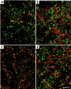
Clustering of GABAA receptors mediated by the endogenous γ3 subunit. The γ3 subunit in the RTN promotes gephyrin and GABAA receptor α3 subunit clustering in γ20/0 mice. Parasagittal sections through the RTN of P14 mice (n = 4–6 per genotype) were double stained for the α3 and γ3 subunit (a and c; α3 red, γ3 green) or the α3 subunit and gephyrin (b and d; α3 red, gephyrin green) and visualized by confocal microscopy. Yellow puncta in the superimposed red and green images illustrate colocalization of the markers used. In γ20/0 RTN, the α3 subunit IR was colocalized with strong IR for the γ3 subunit (a), as well as for gephyrin (b), whereas in γ2+/+ RTN, there was only a partial colocalization of the α3 subunit IR and the weaker γ3 subunit IR (c), but a prominent colocalization of the gephyrin and α3 subunit IR (d). (Scale bar, 10 μm.)

Similar articles

Cited by

References

    1. Colledge M, Froehner S C. Proc Natl Acad Sci USA. 1998;95:3341–3343. - PMC - PubMed
    1. Kirsch J, Meyer G, Betz H. Mol Cell Neurosci. 1996;8:93–98. - PubMed
    1. Mohler H, Fritschy J-M, Lüscher B, Rudolph U, Benson J, Benke D. In: Ion Channels. Narahashi T, editor. Vol. 4. New York: Plenum; 1996. pp. 89–113. - PubMed
    1. Sieghart W. Pharmacol Rev. 1995;47:181–234. - PubMed
    1. Macdonald R L, Olsen R W. Annu Rev Neurosci. 1994;17:569–602. - PubMed