. 1999 Oct 26;96(22):12866-9.
doi: 10.1073/pnas.96.22.12866.

MEK1 protein kinase inhibition protects against damage resulting from focal cerebral ischemia

Affiliations

MEK1 protein kinase inhibition protects against damage resulting from focal cerebral ischemia

A Alessandrini et al. Proc Natl Acad Sci U S A. .

Abstract

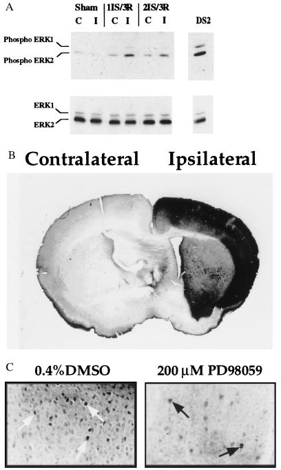
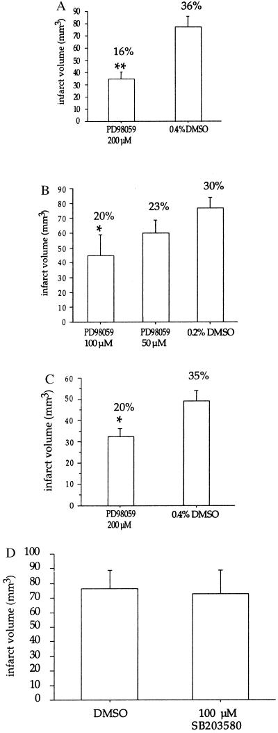
The MEK1 (MAP kinase/ERK kinase)/ERK (extracellular-signal-responsive kinase) pathway has been implicated in cell growth and differentiation [Seger, R. & Krebs, E. G. (1995) FASEB J. 9, 726-735]. Here we show that the MEK/ERK pathway is activated during focal cerebral ischemia and may play a role in inducing damage. Treatment of mice 30 min before ischemia with the MEK1-specific inhibitor PD98059 [Alessi, D. R., Cuenda, A., Cohen, P. , Dudley, D. T. & Saltiel, A. R. (1995) J. Biol. Chem. 270, 27489-27494] reduces focal infarct volume at 22 hr after ischemia by 55% after transient occlusion of the middle cerebral artery. This is accompanied by a reduction in phospho-ERK1/2 immunohistochemical staining. MEK1 inhibition also results in reduced brain damage 72 hr after ischemia, with focal infarct volume reduced by 36%. This study indicates that the MEK1/ERK pathway contributes to brain injury during focal cerebral ischemia and that PD98059, a MEK1-specific antagonist, is a potent neuroprotective agent.


Figures

Figure 1
Figure 1
(A) Increase in ERK1/2 phosphorylation after ischemia and reperfusion. Western blot analysis was performed on lysates (40 μg per lane) from the contralateral (C) and ipsilateral/ischemic (I) side of the brain after 1 hr of ischemia (1IS) followed by 3 min of reperfusion (3R) or 2 hr of ischemia (2IS) followed by 3 min of reperfusion (3R). Western analysis was performed with phospho-specific ERK1/2 antibodies (New England Biolabs) (1:1000 dilution) (Upper). The blot was stripped and reprobed with C-14 ERK2-specific antibodies (Santa Cruz Biotechnology) (1:1000 dilution) (Lower). DS2 total cell lysate (40 μg) was used as a control for ERK1/2 phosphorylation. DS2 is a clonal cell line that exhibits constitutively active ERK1/2 (23). (B) Increased ERK1/2 phosphorylation in the cortical region of the brain after 1 hr of focal cerebral ischemia and 3 min of reperfusion. Immunohistochemistry of brain sections (40 μm thick) was performed with phospho-specific ERK1/2 antibodies. Contralateral (nonischemic) and ipsilateral (ischemic) sides of the brain are indicated. (×100.) (C) Pretreatment of mice with PD98059 leads to decreased ERK1/2 phosphorylation in the cortical region of the brain after 2 hr of focal cerebral ischemia and 3 min of reperfusion. Immunohistochemistry of brain sections (40 μm thick) was performed with the phospho-specific ERK1/2 antibodies. Nuclear staining is indicated by the arrows. (×400.) Dunnett’s posthoc tests. (C) PD98059 neuroprotection is maintained 3 days after ischemia. Brain sections were analyzed as described in the text. Numbers reflect the values from seven mice per treatment; ∗, P < 0.05. The data were analyzed by Student’s t test. Note: The number above each bar reflects the mean infarct volume as a percentage of the contralateral hemisphere to correct for edema, and was calculated using the following formula: (contralateral volume − ipsilateral undamaged volume) × 100/contralateral volume (8). (D) Pretreatment with SB203580, an inhibitor of p38 MAP kinase, is not neuroprotective. Mice were pretreated with 2 μl of 100 μM SB203580 or 0.2% DMSO 30 min before 2 hr of focal cerebral ischemia followed by 22 hr of reperfusion. The numbers reflect the values from four mice per treatment.
Figure 2
Figure 2
Pretreatment with PD98059 is neuroprotective. (A) Mice were pretreated with 200 μM PD98059 or 0.4% DMSO 30 min before 2 hr of focal cerebral ischemia followed by 22 hr of reperfusion. Brains were removed and sliced into five coronal sections (2 mm thick). The sections were treated with 2% 2,3,5-triphenyltetrazolium chloride (Sigma), followed by 10% formalin overnight. The infarcted areas were measured by an image analysis system (MCID version 3) on the posterior surface of each section, and infarction volume was calculated by summing the infarction volume of sequential 2-mm-thick sections. Numbers reflect the values obtained from seven mice per treatment (±SEM); ∗∗, P < 0.01. The data were analyzed by Student’s t test. (B) Dose-dependent neuroprotection with PD98059. Mice were pretreated with 50 or 100 μM PD98059 or 0.2% DMSO 30 min before 2 hr of focal cerebral ischemia followed by 22 hr of reperfusion. Infarcted volume values were obtained as described for A. The numbers reflect the values from seven mice per treatment; ∗, P < 0.05. The data were analyzed by ANOVA followed by

References

    1. Seger R, Krebs E G. FASEB J. 1995;9:726–735. - PubMed
    1. Kindy M S. J Cereb Blood Flow Metab. 1993;13:372–377. - PubMed
    1. Campos-Gonzalez R, Glenney J R., Jr Cell Regul. 1991;2:663–673. - PMC - PubMed
    1. Young S W, Poole R C, Hudson A T, Halestrap A P, Denton R M, Tavare J M. FEBS Lett. 1993;316:278–282. - PubMed
    1. Murray B, Alessandrini A, Cole A J, Yee A G, Furshpan E J. Proc Natl Acad Sci USA. 1998;95:11975–11980. - PMC - PubMed

Publication types

MeSH terms

LinkOut - more resources