. 1999 Oct 26;96(22):12923-8.
doi: 10.1073/pnas.96.22.12923.

Compromised disease resistance in saponin-deficient plants

Affiliations

Compromised disease resistance in saponin-deficient plants

K Papadopoulou et al. Proc Natl Acad Sci U S A. .

Abstract

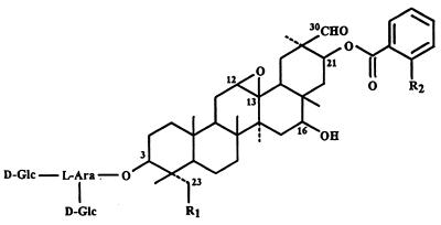
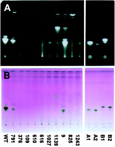
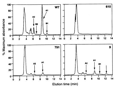
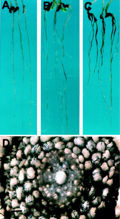
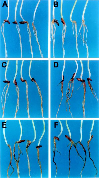
Saponins are glycosylated plant secondary metabolites found in many major food crops [Price, K. R., Johnson, I. T. & Fenwick, G. R. (1987) CRC Crit. Rev. Food Sci. Nutr. 26, 27-133]. Because many saponins have potent antifungal properties and are present in healthy plants in high concentrations, these molecules may act as preformed chemical barriers to fungal attack. The isolation of plant mutants defective in saponin biosynthesis represents a powerful strategy for evaluating the importance of these compounds in plant defense. The oat root saponin avenacin A-1 fluoresces under ultraviolet illumination [Crombie, L., Crombie, W. M. L. & Whiting, D. A. (1986) J. Chem. Soc. Perkins 1, 1917-1922], a property that is extremely rare among saponins. Here we have exploited this fluorescence to isolate saponin-deficient (sad) mutants of a diploid oat species, Avena strigosa. These sad mutants are compromised in their resistance to a variety of fungal pathogens, and a number of lines of evidence suggest that this compromised disease resistance is a direct consequence of saponin deficiency. Because saponins are widespread throughout the plant kingdom, this group of secondary metabolites may have general significance as antimicrobial phytoprotectants.


Figures

Figure 1
Figure 1
The structures of the four avenacins. Avenacin A-1: R1 = OH, R2 = NHCH3; avenacin A-2: R1 = OH, R2 = H; avenacin B-1: R1 = H, R2 =NHCH3; avenacin B-2: R1 = H, R2 = H; d-Glc = d-glucose; l-Ara = l-arabinose.
Figure 2
Figure 2
TLC analysis of partially purified root extracts from wild-type and mutant oats. The fluorescent avenacins, A-1 and B-1, were visualized under UV illumination (Upper), and all four saponins were detected using a chromogenic reagent (Lower). Preparations enriched for each of the four avenacins (3 μg per lane) are on the Right, and extract from wild-type (WT) oat roots is on the Left. The major fluorescent compound present in the wild-type root extract is avenacin A-1, and the minor one with a higher Rf value is avenacin B-1.
Figure 3
Figure 3
HPLC analysis of crude extracts from wild-type and mutant oat roots. The peaks corresponding to the four avenacins are indicated for extracts from wild-type (WT) roots (Upper Left); the peaks eluting at around 5 min derive from micelles of the four avenacins, which are resolved into the separate peaks on re-running. Extracts from 376, 109, 610, 616, 1,027, 1,139, 825, and 1,243 did not contain detectable levels of any of the four avenacins (an example of an HPLC trace for 610 is shown), whereas those from 791 and 9 contained reduced levels (Lower). Extracts from 9 and 1,139 both had an additional peak with a retention time of 12 min, which is likely to correspond to monodeglucosyl avenacin A-1 (indicated for mutant 9 by the unlabeled arrow Lower Right). The full-scale deflection was 4.1 mV for the wild type and 5.5–8.5 mV for the mutants.
Figure 4
Figure 4
Infection of sad mutants by Gaemannomyces graminis var. tritici. (A) A. strigosa wild type; (B and C) sad2 mutants 791 and 1027, respectively (21 days after inoculation); (D) cross-section of an infected root of mutant 1027 viewed under bright field illumination after staining with trypan blue/lactophenol. (Bar = 6 μm.)
Figure 5
Figure 5
Susceptibility of the wild-type A. strigosa line and mutant 610 to Fusarium spp. (A, C, and E) Wild-type seedlings; (B, D, and F) sad mutant 610. (A and B) Mock-inoculated; (C and D) inoculated with Fusarium culmorum isolate F712; (E and F) inoculated with Fusarium avenaceum isolate F616 (assessed after 15 days).

References

    1. Schönbeck F, Schlösser E. In: Physiological Plant Pathology. Heitefuss R, Williams P H, editors. Berlin: Springer; 1976. pp. 653–678.
    1. VanEtten H D, Mansfield J W, Bailey J A, Farmer E E. Plant Cell. 1994;6:1191–1192. - PMC - PubMed
    1. Osbourn A E. Plant Cell. 1996;8:1821–1831. - PMC - PubMed
    1. Müller K O, Börger H. Arb Biol Reichsasnstalt Landw Forstw Berlin. 1940;23:189–223.
    1. Paxton J D. Phytopathol Z. 1981;101:106–109.

LinkOut - more resources