. 1999 Nov;140(11):5310-21.
doi: 10.1210/endo.140.11.7148.

Differential uterine expression of estrogen and progesterone receptors correlates with uterine preparation for implantation and decidualization in the mouse

Affiliations

Differential uterine expression of estrogen and progesterone receptors correlates with uterine preparation for implantation and decidualization in the mouse

J Tan et al. Endocrinology. 1999 Nov.

Abstract

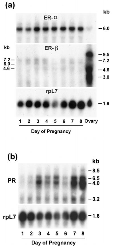
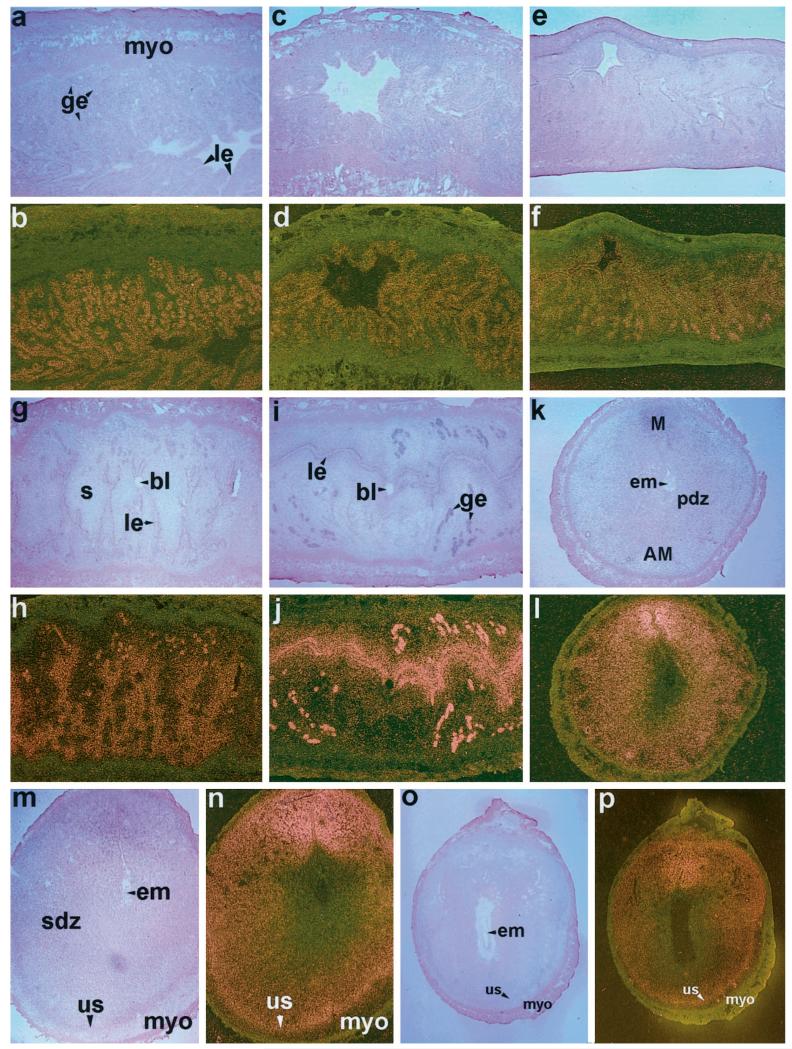
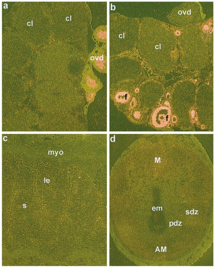
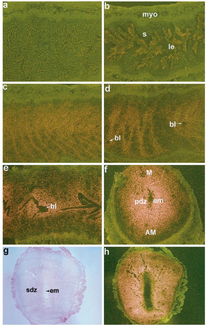
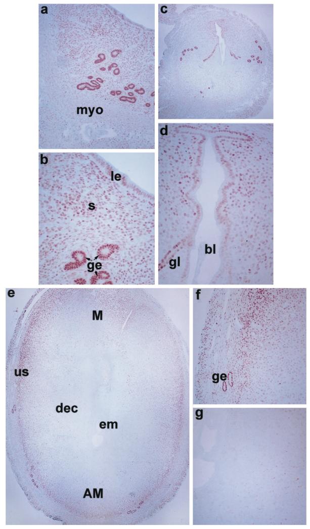
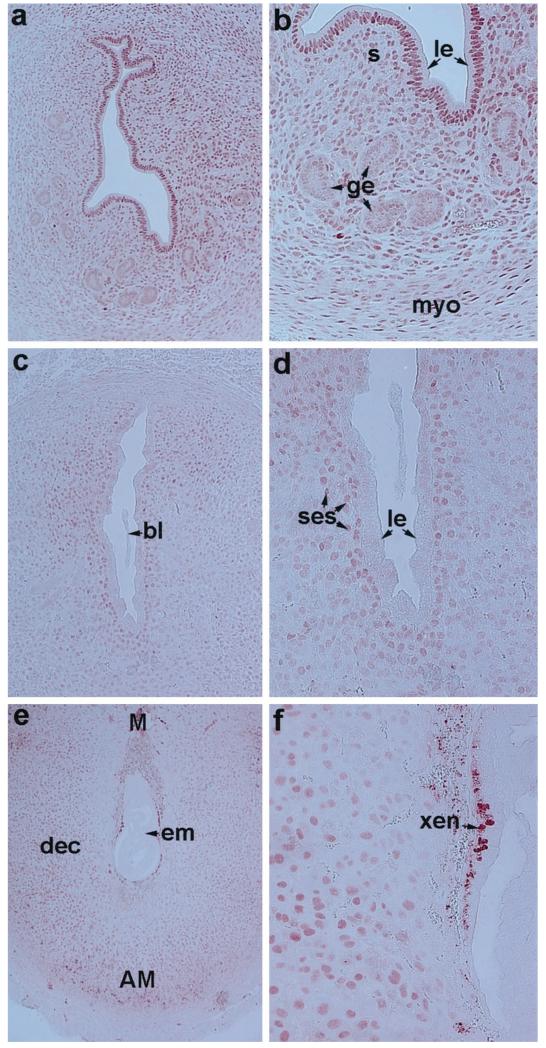
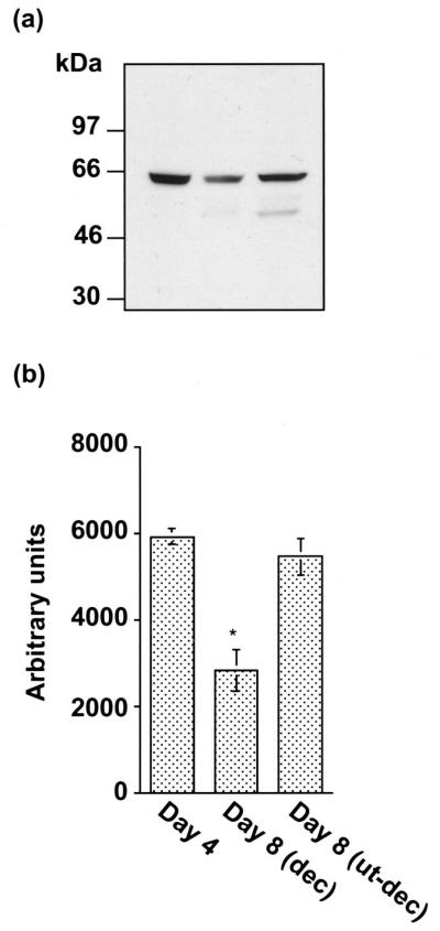
The present investigation examined the spatiotemporal expression of estrogen receptors (ER-alpha and ER-beta) and progesterone receptor (PR) in the periimplantation mouse uterus (days 1-8). ER-alpha messenger RNA (mRNA) was detected at much higher levels in the periimplantation uterus compared with that of ER-beta mRNA, the levels of which were very low in all uterine cells during this period. Results of in situ hybridization demonstrated expression of ER-alpha mRNA primarily in the luminal and glandular epithelia on days 1 and 2 of pregnancy. On days 3 and 4, the accumulation was localized primarily in stromal cells in addition to its presence in the epithelium. Following implantation on day 5, the accumulation of this mRNA was more condensed in the luminal and glandular epithelia, but declined in the subluminal epithelial stroma at the sites of implanting embryos. On days 6-8, the accumulation of ER-alpha mRNA was primarily localized in the secondary decidual zone (SDZ) with more intense localization in the subepithelial cells at the mesometrial pole. In contrast, signals were very low to undetectable in the primary decidual zone (PDZ), and no signals were detected in implanting embryos. The undifferentiated stroma underneath the myometrium also showed positive signals. The immunolocalization of ER-alpha protein correlated with the mRNA localization. Western blot analysis showed down-regulation of ER-alpha in day 8 decidual cell extracts consistent with the down-regulation of ER-alpha mRNA in decidual cells immediately surrounding the embryo on this day. The expression pattern of PR was also dynamic in the periimplantation uterus. On day 1, the accumulation of PR mRNA was very low to undetectable, whereas only a modest level of accumulation in the epithelium was noted on day 2. On days 3 and 4, the accumulation of this mRNA was detected in both the epithelium and stroma. In contrast, the expression was restricted only to the stroma with increased signals at the sites of implantation on day 5. On days 6-8, PR mRNA accumulation increased dramatically throughout the deciduum. The localization of immunoreactive PR correlated with the mRNA distribution in the periimplantation uterus. Taken together, the results demonstrate that the expression of ER-alpha, ER-beta, and PR is differentially regulated in the periimplantation mouse uterus. This compartmentalized expression of ER and PR provides information regarding the sites of coordinated effects ofestrogen and progesterone in the preparation of the uterus for implantation and decidualization during early pregnancy.


Figures

FIG. 1
FIG. 1
a, Northern blot hybridization of ER-α, ER-β, and rpL7 mRNAs in the periimplantation mouse uterus (days 1–8) and ovary. b, Northern blot hybridization of PR and rpL7 mRNAs in the periimplantation uterus (days 1–8). Poly (A)+ RNA (2 μg) was separated by formaldehyde-agarose gel electrophoresis, transferred and UV cross-linked to nylon membrane, and hybridized as described in Materials and Methods. Hybridization was performed using 32P-labeled cRNA probes sequentially to (a) ER-α, ER-β, and rpL7 or (b) PR and rpL7. Autoradiographic exposures were 2 h for ER-α, 5 days for ER-β, 6 h for PR, and 1.5 h for rpL7.
FIG. 2
FIG. 2
In situ hybridization of ER-α mRNA in the periimplantation mouse uterus. Brightfield and darkfield photomicrographs of representative longitudinal sections of uteri on days 1–5 (a–j) at 40×, and cross-sections of uteri on days 6–7 (k–n) at 40× and day 8 (o, p) at 20× are shown. le, Luminal epithelium; ge, glandular epithelium; myo, myometrium; s, stroma; bl, blastocyst; pdz, primary decidual zone; em, embryo; M, mesometrial pole; AM, antimesometrial pole; sdz, secondary decidual zone; us, undifferentiated stroma. These experiments were repeated three times with similar results. Sections hybridized with 35S-labeled sense probe did not show any positive signals (data not shown).
FIG. 3
FIG. 3
In situ hybridization of ER-α and ER-β mRNAs in the ovary and ER-β mRNA in the uterus on days 4 and 8 of pregnancy. Darkfield photomicrographs of representative ovary section hybridized with cRNA probes for ER-α (a), ER-β (b) are shown at 40×. Darkfield photomicrographs of representative uterine sections on days 4 (c) and 8 (d) of pregnancy hybridized with ER-β probe are shown at 40× and 25×, respectively. cl, Corpus luteum; ovd, oviduct; f, follicles; le, luminal epithelium; s, stroma; myo, myometrium; pdz, primary decidual zone; sdz, secondary decidual zone; em, embryo; M, mesometrial pole; AM, antimesometrial pole. These experiments were repeated three times with similar results. Sections hybridized with 35S-labeled sense probe did not show any positive signals (data not shown).
FIG. 4
FIG. 4
In situ hybridization of PR mRNA in the uterus on days 1–7 of pregnancy. Darkfield photomicrographs of representative longitudinal sections of uteri on days 1–5 (a–e) at 40 × and cross-section of uterus on day 6 (f) at 40× are shown. Brightfield and darkfield photomicrographs of uterine cross-section on day 7 (g and h, respectively) at 25× are shown. le, Luminal epithelium; s, stroma; myo, myometrium; bl, blastocyst; pdz, primary decidual zone; em, embryo; M, mesometrial pole; AM, antimesometrial pole; sdz, secondary decidual zone. These experiments were repeated three times with similar results. Sections hybridized with 35S-labeled sense probe did not show any positive signals (data not shown).
FIG. 5
FIG. 5
Immunohistochemistry of ER-α in the uterus on days 4, 5, and 8 of pregnancy. Brightfield photomicrographs of representative uterine sections are shown. Red deposits indicate positive nuclear immunostaining for ER-α. a, Day 4, 100×; b, day 4, 200×; c, day 5, 40×; d, day 5, 200×; e, day 8, 40×; f and g, two areas of (e) at 100×. myo, Myometrium; le, luminal epithelium; ge, glandular epithelium; s, stroma; bl, blastocyst; em, embryo; gl, gland; dec, decidual cells; us, undifferentiated stroma; M, mesometrial pole; AM, antimesometrial pole. These experiments were repeated three times with similar results.
FIG. 6
FIG. 6
Immunohistochemistry of PR in the periimplantation mouse uterus. Brightfield photomicrographs of representative uterine sections are shown. Red deposits indicate positive nuclear immunostaining for PR. a, Day 4, 100×; b, day 4, 200×; c, day 5, 100×; d, day 5, 200×; (e) day 8, 40×; and (f) day 8, 200×. le, Luminal epithelium; ge, glandular epithelium; s, stroma; myo, myometrium; bl, blastocyst; em, embryo; ses, subepithelial stroma; dec, decidual cells; M, mesometrial pole; AM, antimesometrial pole; xen, extraembryonic endoderm. These experiments were repeated three times with similar results.
FIG. 7
FIG. 7
a, Western blot analysis of ER-α in extracts of whole uterus on day 4 of pregnancy, and the separated decidua and the uterus minus decidua on day 8 of pregnancy. Immunoblotting with the primary antibody for ER-α was performed as described in Materials and Methods. b, The quantitation of ER-α protein levels was examined by densitometric scanning (Personal Densitometer SI, Molecular Dynamics, Inc., Sunnyvale, CA) of the immunoblots. Results are mean ± SEM from three independent experiments. *, Significantly different from other groups (P < 0.05).

References

    1. Martin L, Finn CA, Trinder G. Hypertrophy and hyperplasia in the mouse uterus after estrogen treatment: an autoradiographic study. J Endocrinol. 1973;56:133–144. - PubMed
    1. Huet-Hudson YM, Andrews GK, Dey SK. Cell type-specific localization of c-Myc protein in the mouse uterus: modulation by steroid hormones and analysis of the periimplantation period. Endocrinology. 1989;125:1683–1690. - PubMed
    1. Martin L, Das RM, Finn CA. The inhibition by progesterone of uterine epithelial proliferation in the mouse. J Endocrinol. 1973;57:549–554. - PubMed
    1. Psychoyos A. Endocrine control of egg implantation. In: Greep RO, Astwood EG, Geiger SR, editors. Handbook of Physiology. American Physiological Society; Washington DC: 1973. pp. 187–215.
    1. Paria BC, Huet-Hudson YM, Dey SK. Blastocyst’s state of activity determines the “window” of implantation in the receptive mouse uterus. Proc Natl Acad Sci USA. 1993;90:10159–10162. - PMC - PubMed

Publication types