

CCR5 and CXCR4 chemokine receptor expression and beta-chemokine production during early T cell repopulation induced by highly active anti-retroviral therapy
- PMID: 10540164
- PMCID: PMC1905399
- DOI: 10.1046/j.1365-2249.1999.01033.x
CCR5 and CXCR4 chemokine receptor expression and beta-chemokine production during early T cell repopulation induced by highly active anti-retroviral therapy
Abstract
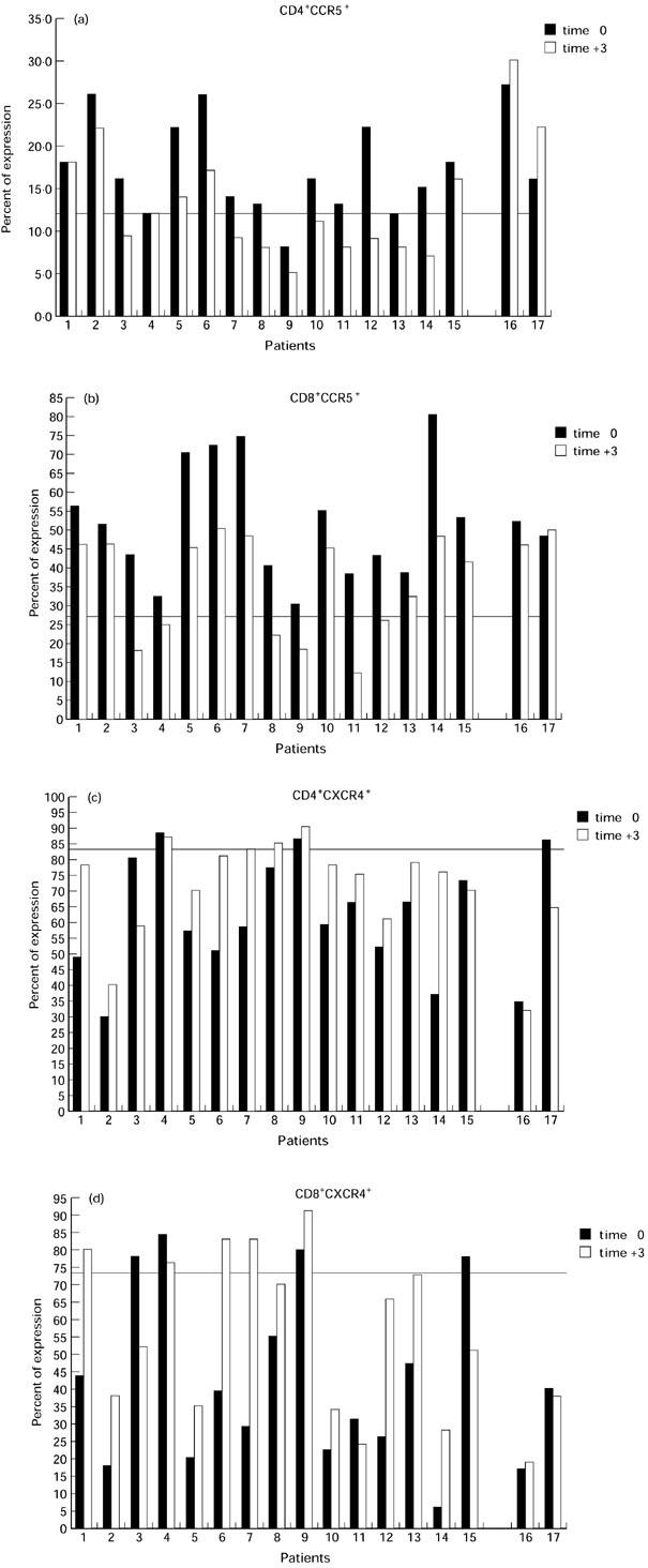
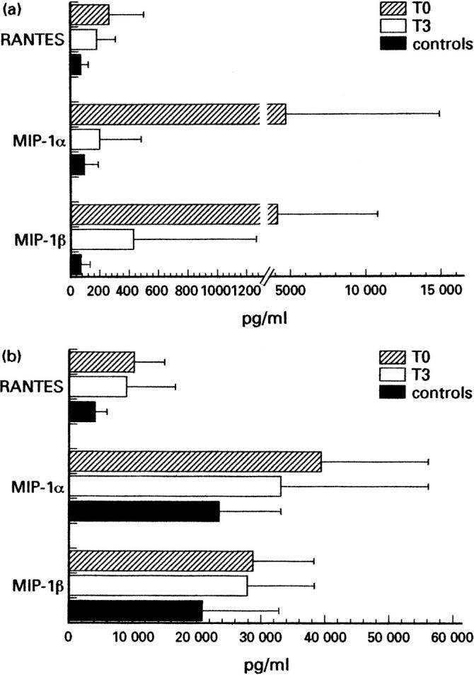
Expression of chemokine receptors and beta-chemokine production by peripheral blood mononuclear cells (PBMC) were determined in HIV-1-infected individuals before and after highly active anti-retroviral therapy (HAART) and their relationship to viral load, T cell phenotype and the expression of immunological activation markers was examined. We found that the expression of CCR5 is up-regulated in HIV-1-infected individuals while CXCR4 appears down-regulated on both CD4 and CD8 T cells compared with normal controls. These alterations are associated with the high levels of viral load. In addition, a relationship was observed between the degree of immune activation and chemokine receptor expression on T cells. However, after 3 months of combined anti-retroviral regimen, expression of CXCR4 significantly increased while CCR5 decreased when compared with pretherapy determinations. This was seen in strict association with a dramatic decrease of viral load and an increase of both CD45RA+/CD62L+ (naive) and CD45RA-/CD62L+ or CD45RA+/CD62L- (memory) T cells accompanied by a significant decrease of the expression of immune activation markers such as HLA-DR and CD38. At enrolment, both spontaneous and lectin-induced RANTES, macrophage inflammatory protein-1alpha (MIP-1alpha) and MIP-1beta production by PBMC were higher in HIV-1-infected individuals compared with normal controls, although differences for MIP-1beta were not statistically significant. However, RANTES and MIP-1alpha production decreased during HAART at levels closer to that determined with normal controls, while MIP-1beta production was less consistently modified. These data indicate that the expression of chemokine receptors CCR5 and CXCR4 and the production of beta-chemokines are altered in HIV-infected individuals, and suggest that their early modifications during HAART reflect both the peripheral redistribution of naive/memory T cell compartments and the decrease in levels of T cell activation. Such modifications in the expression of host determinants of viral tropism and the production of anti-viral molecules may play a role in the emergence of virus variants when a failure of HAART occurs.
Figures
References
-
- Dragic T, Litwin V, Allaway GP, et al. HIV-1 entry into CD4+ cells is mediated by the chemokine receptor CC- CKR-5. Nature. 1996;381:667–73. - PubMed
-
- Deng H, Liu R, Ellmeier W, et al. Identification of a major co-receptor for primary isolates of HIV-1. Nature. 1996;381:661–6. - PubMed
-
- Choe H, Farzan M, Sun Y, et al. The beta-chemokine receptors CCR3 and CCR5 facilitate infection by primary HIV-1 isolates. Cell. 1996;85:1135–48. - PubMed
-
- Carroll RG, Riley JL, Levine BL, Blair PJ, St LD, June CH. The role of co-stimulation in regulation of chemokine receptor expression and HIV-1 infection in primary T lymphocytes. Semin Immunol. 1998;10:195–202. - PubMed
-
- Feng Y, Broder CC, Kennedy PE, Berger EA. HIV-1 entry cofactor: functional cDNA cloning of a seven-transmembrane, G protein-coupled receptor. Science. 1996;272:872–7. - PubMed
Publication types
MeSH terms
Substances
LinkOut - more resources
Full Text Sources
Other Literature Sources
Medical
Research Materials
