

Cytolytic mechanisms involved in non-MHC-restricted cytotoxicity in Chediak-Higashi syndrome
- PMID: 10540167
- PMCID: PMC1905392
- DOI: 10.1046/j.1365-2249.1999.01025.x
Cytolytic mechanisms involved in non-MHC-restricted cytotoxicity in Chediak-Higashi syndrome
Abstract
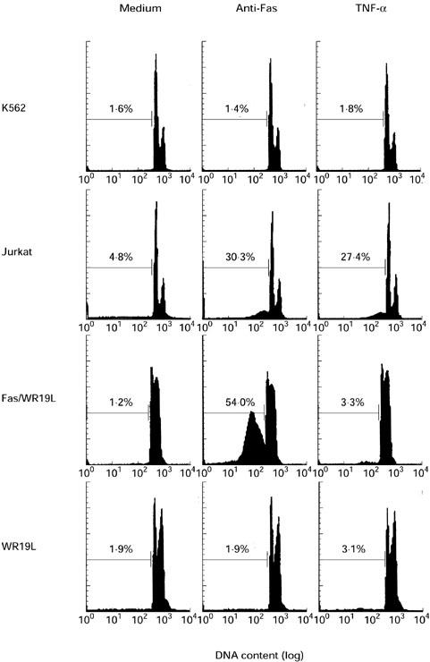
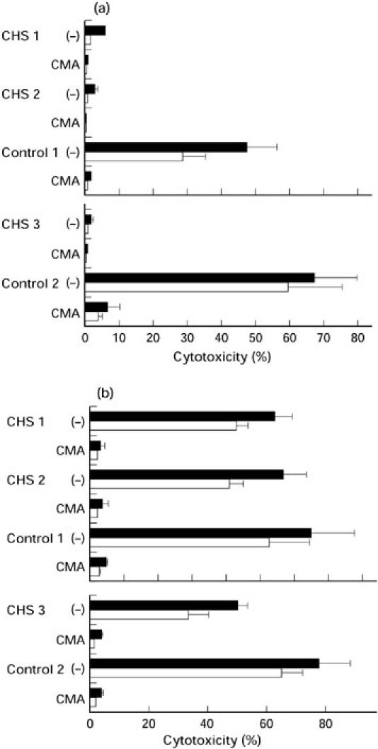
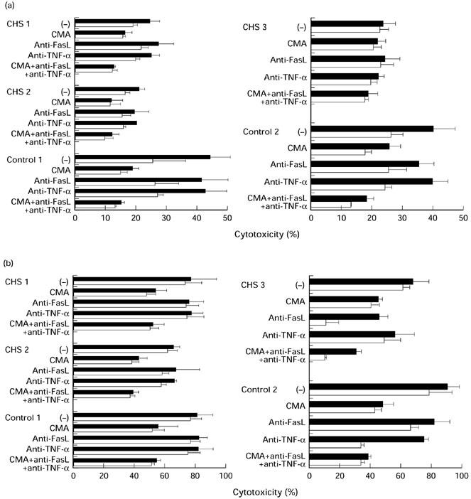
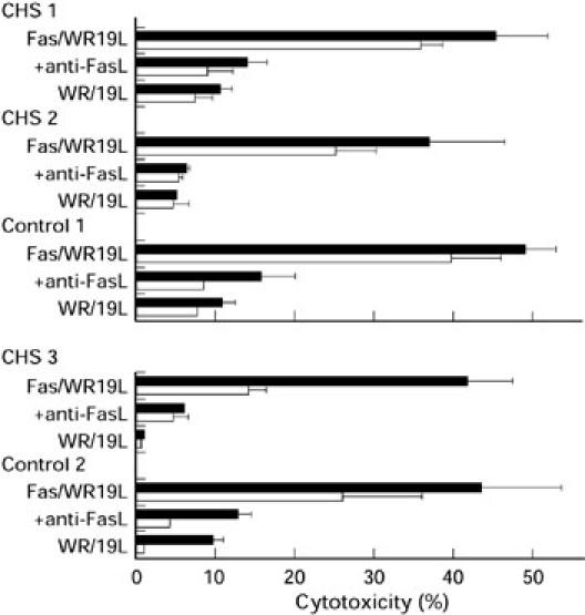
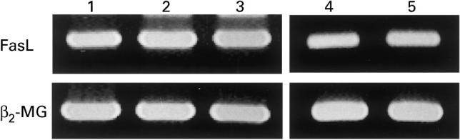
To determine the mechanisms responsible for the impaired lymphocyte-mediated cytotoxicity in Chediak-Higashi syndrome (CHS), we investigated the killing ability of peripheral blood lymphocytes (PBL) from three patients with CHS using several kinds of target cells that were sensitive to perforin, Fas ligand (FasL), and/or tumour necrosis factor-alpha (TNF-alpha). Freshly isolated CHS PBL did not kill K562 target cells, killing of which by normal PBL was perforin-dependent, as demonstrated by complete inhibition by concanamycin A (CMA), an inhibitor of perforin-based cytotoxicity. In contrast, the CHS PBL exhibited substantial cytotoxicity against Jurkat cells, which was only partially inhibited by CMA treatment but not by the addition of neutralizing anti-FasL or anti-TNF-alpha antibodies. IL-2-activated CHS PBL exhibited substantial levels of cytotoxicity against K562 and Jurkat cells, the levels being 74% and 83% of the respective normal control values, respectively. CMA treatment showed that while the cytotoxicity of IL-2-activated CHS PBL against K562 was largely dependent on perforin, that against Jurkat was largely not. IL-2-activated CHS PBL expressed FasL mRNA, and killed Fas transfectants. These findings indicate that CHS PBL have an ability to kill some target cells via a perforin-mediated pathway, especially when they are activated by IL-2. It was also demonstrated that CHS PBL can exert cytotoxicity against certain target cells by utilizing FasL and an undefined effector molecule other than perforin, FasL, or TNF-alpha.
Figures
Similar articles
-
Enhancement of human cord blood CD34+ cell-derived NK cell cytotoxicity by dendritic cells.J Immunol. 2001 Feb 1;166(3):1590-600. doi: 10.4049/jimmunol.166.3.1590. J Immunol. 2001. PMID: 11160200
-
Interferon-alpha (IFN-alpha) enhances cytotoxicity in healthy volunteers and chronic hepatitis C infection mainly by the perforin pathway.Clin Exp Immunol. 1999 Oct;118(1):71-7. doi: 10.1046/j.1365-2249.1999.01020.x. Clin Exp Immunol. 1999. PMID: 10540162 Free PMC article.
-
Perforin-mediated lysis of tumor cells by Mycobacterium bovis Bacillus Calmette-Guérin-activated killer cells.Clin Cancer Res. 2000 Sep;6(9):3729-38. Clin Cancer Res. 2000. PMID: 10999767
-
Do macrophages kill through apoptosis?Immunol Today. 1996 Dec;17(12):573-6. doi: 10.1016/s0167-5699(96)10071-2. Immunol Today. 1996. PMID: 8991289 Review.
-
The role of cytotoxicity in lymphocyte homeostasis.Curr Opin Immunol. 2001 Oct;13(5):549-54. doi: 10.1016/s0952-7915(00)00257-0. Curr Opin Immunol. 2001. PMID: 11544002 Review.
Cited by
-
Involvement of CD27/CD70 interactions in antigen-specific cytotoxic T-lymphocyte (CTL) activity by perforin-mediated cytotoxicity.Clin Exp Immunol. 2002 Dec;130(3):424-30. doi: 10.1046/j.1365-2249.2002.02012.x. Clin Exp Immunol. 2002. PMID: 12452832 Free PMC article.
References
-
- Chediak M. Nouvelle anomalie leukocytaire de caractere constitutionnel et familiel. Rev Hematol. 1952;7:362–7. - PubMed
-
- Higashi O. Congenital gigantism of peroxidase granules. Tohoku J Exp Med. 1954;59:315–32. - PubMed
-
- Blume RS, Wolff SM. The Chediak-Higashi syndrome: studies in four patients and a review of the literature. Medicine. 1972;51:247–80. - PubMed
-
- Barak Y, Nir E. Chediak-Higashi syndrome. Am J Pediatr Hematol Oncol. 1987;9:42–55. - PubMed
-
- White JG. The Chediak-Higash syndrome: a possible lysosomal disease. Blood. 1966;28:143–56. - PubMed
MeSH terms
Substances
LinkOut - more resources
Full Text Sources
Other Literature Sources
Research Materials
Miscellaneous
