

CD4 cytotoxic and dendritic cells in the immunopathologic lesion of Sjögren's syndrome
- PMID: 10540173
- PMCID: PMC1905402
- DOI: 10.1046/j.1365-2249.1999.01037.x
CD4 cytotoxic and dendritic cells in the immunopathologic lesion of Sjögren's syndrome
Abstract
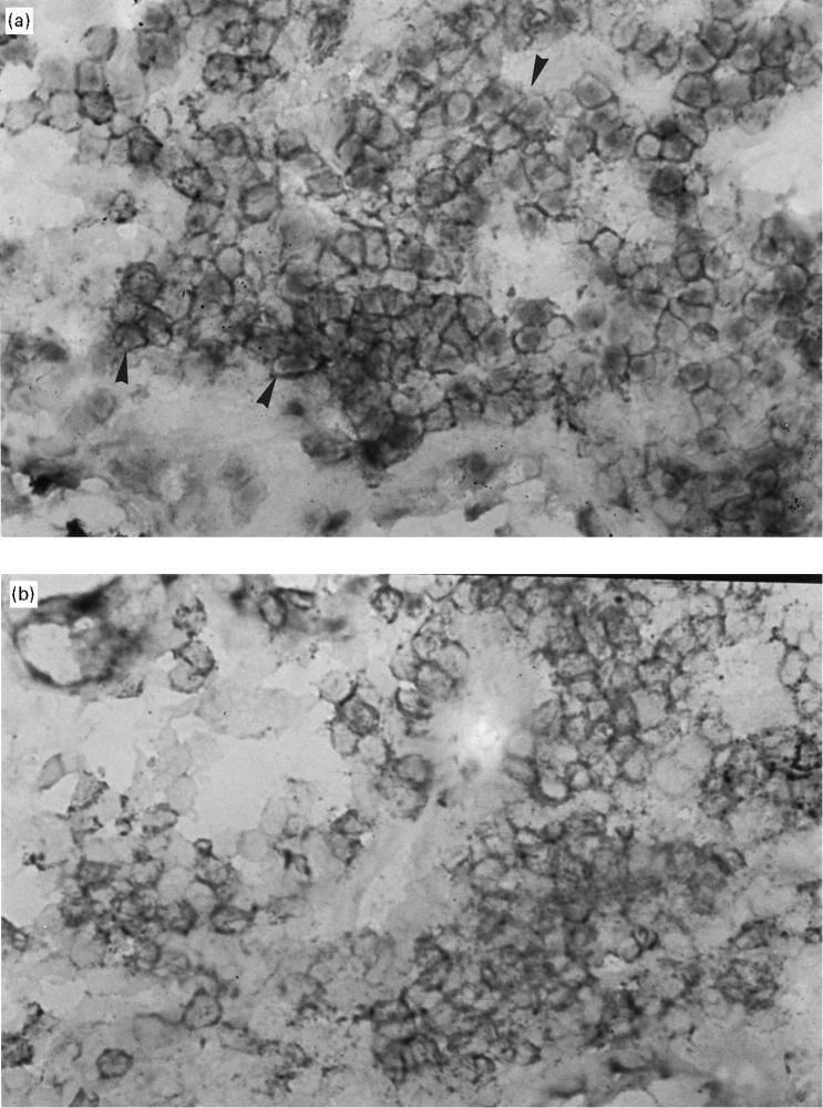
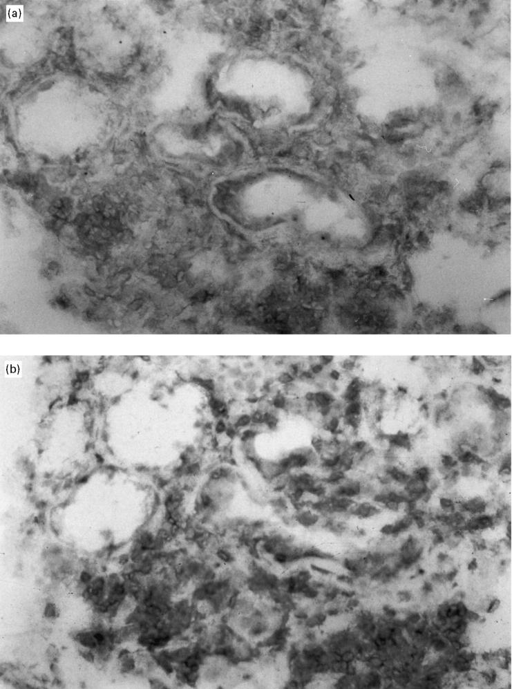
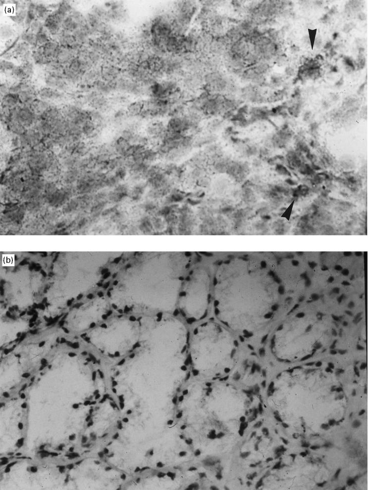
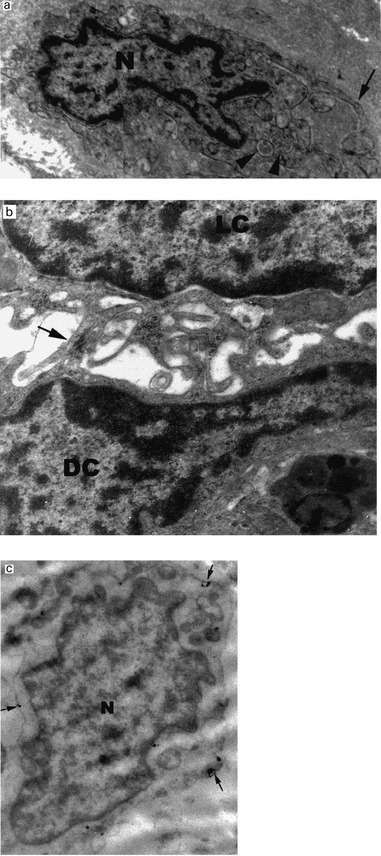
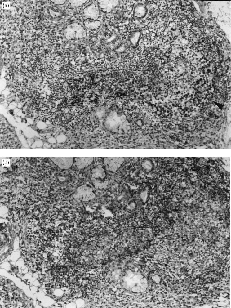
The existence of CD4+ T lymphocytes with cytotoxic activity in minor salivary gland (MSG) biopsies from Sjögren's syndrome (SS) patients was investigated using in situ double immunohistochemistry technique. The presence of dendritic cells (DC) in SS lesions was examined by using single and double immunohistochemistry methods and a panel of different MoAbs to specific cell surface markers (i.e. CD3, CD11c, DRC). Furthermore, the ultrastructural morphology of DC was characterized by electron microscopy (EM). Immunogold labelling technique using the DRC surface marker was also applied. Finally, we investigated the existence of germinal centres (GC) in the salivary gland lesions of SS patients. Seven patients with primary SS and five patients with non-specific sialadenitis were the subjects of this study. Our results indicate the existence of a CD4+ cytotoxic cell population that utilizes perforin-mediated cell destructions as they expressed perforin mRNA. Quantitative analysis of these cells revealed that they comprised approximately 20% of the existing T lymphocytes. We also identified a population of CD4+ T cells that expressed the CD11c activation marker. Furthermore, we observed a distinct cell subtype which expressed the DRC cell surface marker. These cells had the characteristic ultrastructural morphology of DC and were DRC+ when examined by immunoelectron microscopy. Finally, the formation of GC structures in the histopathologic lesions of the salivary glands was observed. The above findings indicate that both CD4+ cytotoxic T lymphocytes (CTL) and DC may be involved in the initiation and perpetuation of SS pathogenesis. Moreover, the formation of GC in the lesions reveals a possible mechanism for in situ differentiation and proliferation of activated B lymphocytes.
Figures
Similar articles
-
Cellular basis of ectopic germinal center formation and autoantibody production in the target organ of patients with Sjögren's syndrome.Arthritis Rheum. 2003 Nov;48(11):3187-201. doi: 10.1002/art.11311. Arthritis Rheum. 2003. PMID: 14613282
-
The Significance of Ectopic Germinal Centers in the Minor Salivary Gland of Patients with Sjögren's Syndrome.J Korean Med Sci. 2016 Feb;31(2):190-5. doi: 10.3346/jkms.2016.31.2.190. Epub 2016 Jan 13. J Korean Med Sci. 2016. PMID: 26839471 Free PMC article.
-
Inflammatory Stratification in Primary Sjögren's Syndrome Reveals Novel Immune Cell Alterations in Patients' Minor Salivary Glands.Front Immunol. 2021 Jul 12;12:701581. doi: 10.3389/fimmu.2021.701581. eCollection 2021. Front Immunol. 2021. PMID: 34322130 Free PMC article.
-
Labial salivary gland biopsy in Sjögren's syndrome.Neth J Med. 1992 Apr;40(3-4):148-57. Neth J Med. 1992. PMID: 1603205 Review.
-
Sjögren's syndrome: pathogenesis.Eur J Ophthalmol. 1999 Jan-Mar;9(1):1-7. Eur J Ophthalmol. 1999. PMID: 10230585 Review.
Cited by
-
Dynamic ocular surface and lacrimal gland changes induced in experimental murine dry eye.PLoS One. 2015 Jan 15;10(1):e0115333. doi: 10.1371/journal.pone.0115333. eCollection 2015. PLoS One. 2015. PMID: 25590134 Free PMC article.
-
MicroRNA in Sjögren's Syndrome: Their Potential Roles in Pathogenesis and Diagnosis.J Immunol Res. 2018 Jun 7;2018:7510174. doi: 10.1155/2018/7510174. eCollection 2018. J Immunol Res. 2018. PMID: 29977932 Free PMC article. Review.
-
Pharmacological cholinergic blockade stimulates inflammatory cytokine production and lymphocytic infiltration in the mouse lacrimal gland.Invest Ophthalmol Vis Sci. 2011 May 16;52(6):3221-7. doi: 10.1167/iovs.09-4212. Invest Ophthalmol Vis Sci. 2011. PMID: 21273534 Free PMC article.
-
Impact of interleukin-21 in the pathogenesis of primary Sjögren's syndrome: increased serum levels of interleukin-21 and its expression in the labial salivary glands.Arthritis Res Ther. 2011;13(5):R179. doi: 10.1186/ar3504. Epub 2011 Oct 26. Arthritis Res Ther. 2011. PMID: 22030011 Free PMC article.
-
Local delivery of AAV2-CTLA4IgG decreases sialadenitis and improves gland function in the C57BL/6.NOD-Aec1Aec2 mouse model of Sjögren's syndrome.Arthritis Res Ther. 2012 Feb 27;14(1):R40. doi: 10.1186/ar3753. Arthritis Res Ther. 2012. PMID: 22369699 Free PMC article.
References
-
- Moutsopoulos HM. Immunopathology of Sjögren's syndrome: more questions than answers. Lupus. 1993;2:49–53. - PubMed
-
- Daniels TE, Aufdemorte THB, Greenspan JS. Daniels TE, Tala LN, Moutsopoulos HM, Kassan SS. Sjögren's syndrome. Clinical and immunological aspects. Berlin: Springer; 1987. Histopathology of Sjögren's syndrome; pp. 258–65.
-
- Kong L, Ogawa N, Nakabayashi T, et al. Fas and FasLigand expression in the salivary glands of patients with primary Sjögren's syndrome. Arthritis Rheum. 1997;40:87–97. - PubMed
-
- Golding H, Singer A. Specificity, phenotype and precursor frequency of primary cytolytic T lymphocytes specific for class II major histocompatibility antigens. J Immunol. 1985;135:1610–5. - PubMed
Publication types
MeSH terms
Substances
LinkOut - more resources
Full Text Sources
Other Literature Sources
Medical
Research Materials
Miscellaneous
