

Keratinocytes from patients with lupus erythematosus show enhanced cytotoxicity to ultraviolet radiation and to antibody-mediated cytotoxicity
- PMID: 10540174
- PMCID: PMC1905409
- DOI: 10.1046/j.1365-2249.1999.01026.x
Keratinocytes from patients with lupus erythematosus show enhanced cytotoxicity to ultraviolet radiation and to antibody-mediated cytotoxicity
Abstract
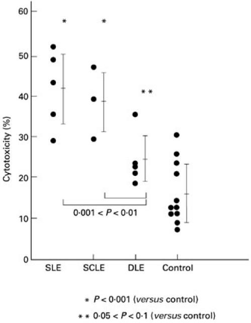
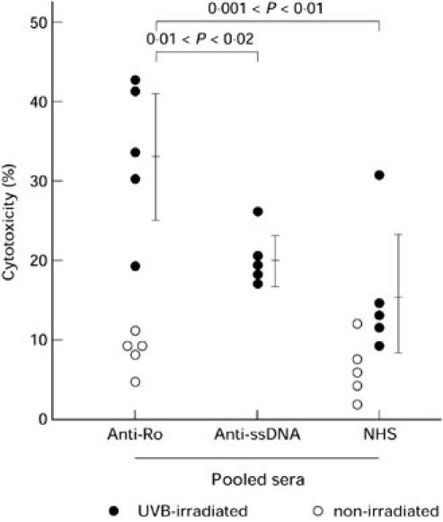
Keratinocyte cytotoxicity is an important component of the immunopathology of photosensitive lupus erythematosus, and antibody-dependent cell-mediated cytotoxicity (ADCC) has been shown to be an important mechanism by which autoantibodies, especially those specific for SS-A/Ro, can induce keratinocyte damage in models of photosensitive lupus. We provide further evidence that keratinocytes from patients with photosensitive lupus show significantly greater ultraviolet radiation (UVR)-induced cytotoxicity, and that ADCC of these targets is especially enhanced by autologous patient's serum or by anti-SS-A/Ro+ sera. Keratinocytes from normal uninvolved skin of 29 patients with cutaneous lupus erythematosus (LE) were grown in cell culture and tested as targets in cytotoxicity experiments in vitro. Cultured keratinocytes from patients with systemic lupus erythematosus (SLE) and subacute cutaneous lupus erythematosus (SCLE) showed significantly greater cytotoxicity following UVR treatment than did keratinocytes from normal adult controls or from neonatal foreskins (P < 0.01). The same cultures also showed greater UVR-induced binding of IgG from fractionated anti-SS-A/Ro+ preparations. ADCC experiments were also performed using keratinocytes cultured from patients with SLE, SCLE, discoid lupus erythematosus (DLE), and normal controls. When keratinocytes were incubated in autologous serum plus a standard mononuclear cell effector population, the percentage of ADCC observed was significantly greater in cultures containing keratinocytes and sera from the SLE and SCLE patients (P < 0.001). When cultured keratinocytes were added to different IgG antibody probes, plus standard mononuclear effector populations, greater ADCC was seen using the anti-SS-A/Ro probe and keratinocytes from patients with SLE or SCLE. With normal human neonatal keratinocyte targets, the anti-SS-A/Ro probe induced greater ADCC than that seen with anti-ssDNA or normal human serum. We have shown that keratinocytes from patients with some forms of lupus erythematosus (SLE and SCLE) show greater cytotoxicity in vitro when irradiated with UVR, and greater susceptibility to ADCC whether the antibody source is their own serum or an anti-SS-A/Ro probe.
Figures
References
-
- Cripps DJ, Rankin J. Action spectra of lupus erythematosus and experimental immunofluorescence. Arch Dermatol. 1973;107:563–7. - PubMed
-
- Freeman RG, Knox JM, Owen DW. Cutaneous lesions of lupus erythematosus induced by monochromatic light. Arch Dermatol. 1969;100:677–82. - PubMed
-
- Walchner M, Messer G, Kind P. Phototesting and photoprotection in LE. Lupus. 1997;6:176–84. - PubMed
-
- Tebbe B, Orfanos CE. Epidemiology and socioeconomic impact of skin disease in lupus erythematosus. Lupus. 1997;6:96–104. - PubMed
-
- Norris DA, Lela LA. Pathogenesis of cutaneous lupus erythematosus. Clin Dermatol. 1985;3:20–35. - PubMed
Publication types
MeSH terms
Substances
Grants and funding
LinkOut - more resources
Full Text Sources
Medical
Research Materials
