. 1999 Sep-Oct;6(5):500-8.
doi: 10.1101/lm.6.5.500.

Brain gene expression during REM sleep depends on prior waking experience

Affiliations

Brain gene expression during REM sleep depends on prior waking experience

S Ribeiro et al. Learn Mem. 1999 Sep-Oct.

Abstract

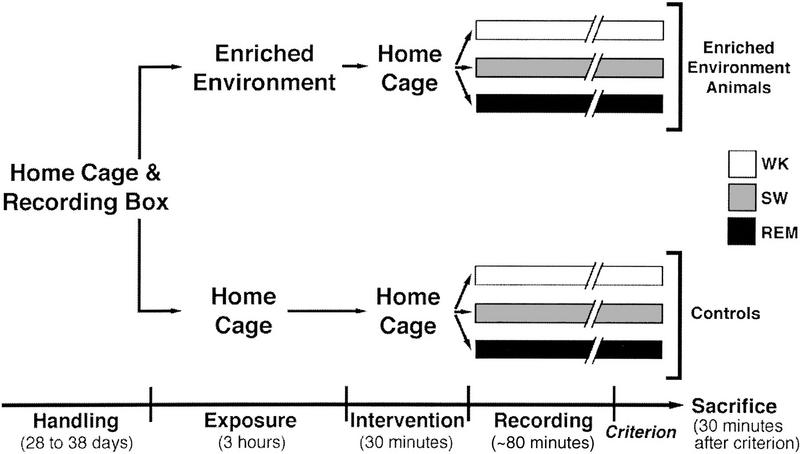
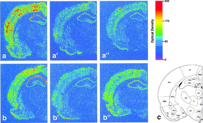
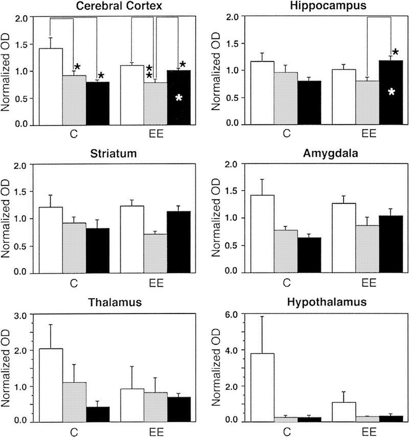
In most mammalian species studied, two distinct and successive phases of sleep, slow wave (SW), and rapid eye movement (REM), can be recognized on the basis of their EEG profiles and associated behaviors. Both phases have been implicated in the offline sensorimotor processing of daytime events, but the molecular mechanisms remain elusive. We studied brain expression of the plasticity-associated immediate-early gene (IEG) zif-268 during SW and REM sleep in rats exposed to rich sensorimotor experience in the preceding waking period. Whereas nonexposed controls show generalized zif-268 down-regulation during SW and REM sleep, zif-268 is upregulated during REM sleep in the cerebral cortex and the hippocampus of exposed animals. We suggest that this phenomenon represents a window of increased neuronal plasticity during REM sleep that follows enriched waking experience.


Figures

Figure 1
Figure 1
Schematic flowchart of the experimental design. All rats were allowed to get familiarized with the home cage, experimenter, and recording conditions for several days, during which baseline EEG was recorded. On the day of the experiment, animals were either exposed to a complex labyrinth (EE) or kept in their home cages (controls) for the last 3 hr of the waking period, followed by an intervening period of 30 min back in the home cages. The animals were then placed in the recording box for monitoring of wake/sleep states until reaching criterion for WK, SW, or REM; the typical duration of this period was ∼80 min. Thirty minutes after criterion, animals were killed and their brains were processed for zif-268 expression by in situ hybridization.
Figure 2
Figure 2
Effect of previous sensorimotor experience on zif-268 brain expression during waking and sleep states. Shown are autoradiograms of brain sections whose gene expression levels best represent the means for each group studied. In controls, zif-268 expression decreased from WK (a) to SW (a′) and REM (a′′). In enriched environment animals, zif-268 levels decreased from WK (b) to SW (b′), but increased from the latter to REM (b′′). This effect was particularly noticeable in the cerebral cortex and the hippocampus. (c) Schematic diagram of a frontal section of the rat brain at the level studied; the 25 regions for which expression was quantified are indicated (for abbreviations see Materials and Methods).
Figure 3
Figure 3
Analysis of zif-268 expression during waking and sleep states in six major brain regions; cerebral cortex, hippocampus, striatum, amygdala, thalamus, and hypothalamus. Statistically significant interactions (MANOVA) between expression profiles occurred for the cerebral cortex (P = 0.04) and the hippocampus (P = 0.03). Whereas zif-268 expression in controls decreased from WK to SW and to REM, in animals exposed to the enriched environment, zif-268 levels decreased from WK to SW but increased from SW to REM. Values on the y-axis represent normalized optical density (OD) measurements (means ± s.e.m.) of X-ray film autoradiograms; statistically significant differences (Bonferroni-Dunn) are indicated by one (P<0.05) or two (P<0.01) asterisks. White, gray, and black columns represent, respectively, WK, SW, and REM. Asterisks inside columns indicate state differences between C and EE groups.
Figure 4
Figure 4
Analysis of zif-268 expression during waking and sleep states in individual brain structures. (Top panels) Three individual structures within the cerebral cortex and the hippocampus in which the most significant differences occurred. Notice that a significant increase in zif-268 expression during REM in EE animals occurred in areas associated with sensory (piriform cortex, P = 0.01), motor (frontal cortex, P = 0.03) and spatial (dentate gyrus, P = 0.03) processing. (Bottom panels) Three individual brain structures in which no significant effect was observed. Ordinates, columns, and asterisks as in Fig. 3.

Similar articles

Cited by

References

    1. Abraham WC, Mason SE, Demmer J, Williams JM, Richardson CL, Tate WP. Correlations between immediate early gene induction and the persistence of long-term potentiation. Neuroscience. 1993;56:717–727. - PubMed
    1. Ambrosini MV, Sadile AG, Gironi CU, Mattiaccio M, Giuditta A. The sequential hypothesis on sleep function. I. Evidence that the structure of sleep depends on the nature of the previous waking experience. Physiol Behav. 1988;43:325–337. - PubMed
    1. Aston-Jones G, Bloom FE. Activity of norepinephrine-containing locus coeruleus neurons in behaving rats anticipates fluctuations in the sleep-waking cycle. J Neurosci. 1981a;1:876–886. - PMC - PubMed
    1. ————— Norepinephrine-containing locus coeruleus neurons in behaving rats exhibit pronounced responses to non-noxious environmental stimuli. J Neurosci. 1981b;1:887–900. - PMC - PubMed
    1. Basheer R, Sherin JE, Saper CB, Morgan JI, McCarley RW, Shiromani PJ. Effects of sleep on wake-induced c-fos expression. J Neurosci. 1997;17:9746–9750. - PMC - PubMed

Publication types

LinkOut - more resources