. 1999 Nov;181(21):6591-9.
doi: 10.1128/JB.181.21.6591-6599.1999.

Functional interactions of a homolog of proliferating cell nuclear antigen with DNA polymerases in Archaea

Affiliations

Functional interactions of a homolog of proliferating cell nuclear antigen with DNA polymerases in Archaea

I K Cann et al. J Bacteriol. 1999 Nov.

Abstract

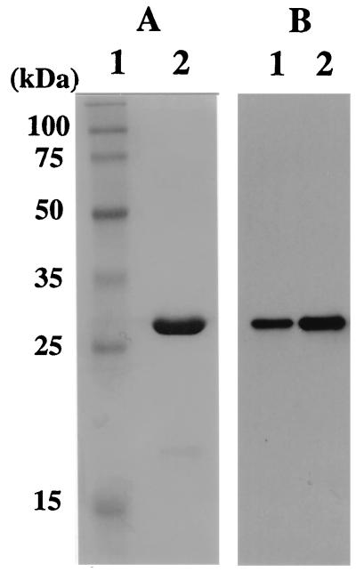
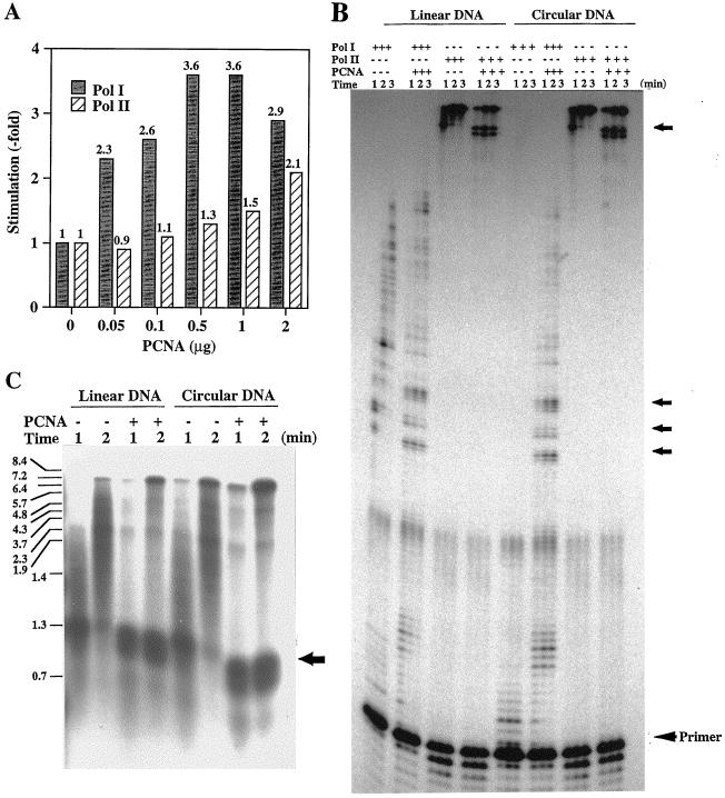
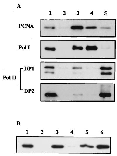
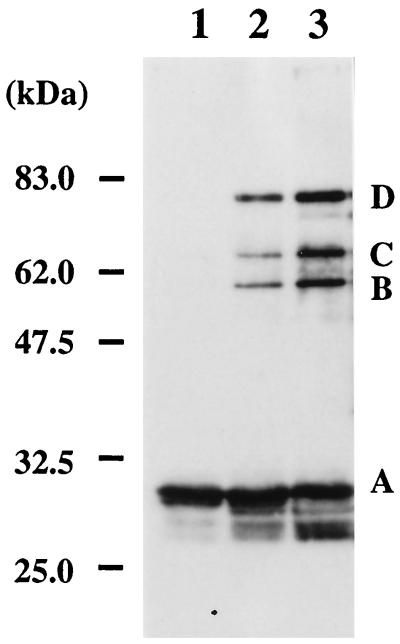
Proliferating cell nuclear antigen (PCNA) is an essential component of the DNA replication and repair machinery in the domain Eucarya. We cloned the gene encoding a PCNA homolog (PfuPCNA) from an euryarchaeote, Pyrococcus furiosus, expressed it in Escherichia coli, and characterized the biochemical properties of the gene product. The protein PfuPCNA stimulated the in vitro primer extension abilities of polymerase (Pol) I and Pol II, which are the two DNA polymerases identified in this organism to date. An immunological experiment showed that PfuPCNA interacts with both Pol I and Pol II. Pol I is a single polypeptide with a sequence similar to that of family B (alpha-like) DNA polymerases, while Pol II is a heterodimer. PfuPCNA interacted with DP2, the catalytic subunit of the heterodimeric complex. These results strongly support the idea that the PCNA homolog works as a sliding clamp of DNA polymerases in P. furiosus, and the basic mechanism for the processive DNA synthesis is conserved in the domains Bacteria, Eucarya, and Archaea. The stimulatory effect of PfuPCNA on the DNA synthesis was observed by using a circular DNA template without the clamp loader (replication factor C [RFC]) in both Pol I and Pol II reactions in contrast to the case of eukaryotic organisms, which are known to require the RFC to open the ring structure of PCNA prior to loading onto a circular DNA. Because RFC homologs have been found in the archaeal genomes, they may permit more efficient stimulation of DNA synthesis by archaeal DNA polymerases in the presence of PCNA. This is the first stage in elucidating the archaeal DNA replication mechanism.


Figures

FIG. 1
FIG. 1
Nucleotide and deduced amino acid sequences of the gene for the P. furiosus PCNA homolog. The BamHI site used for cloning of the structural gene into pET21a and the determined N-terminal amino acid sequence of the truncated protein produced in E. coli are underlined. The asterisk indicates the stop codon.
FIG. 2
FIG. 2
Purification of recombinant PfuPCNA from E. coli cells and identification of PfuPCNA in P. furiosus cells. (A) Recombinant PfuPCNA purified as described in the text was loaded onto an SDS–12.5% polyacrylamide gel and stained with Coomassie brilliant blue. Lanes: 1, molecular mass markers (Perfect Protein Markers; Novagen); 2, purified PfuPCNA (3 μg). The sizes of the molecular mass markers are indicated on the left. (B) Western blot analysis of PfuPCNA. P. furiosus cell extracts (35 μg of cells) and recombinant PCNA (0.01 μg) produced in E. coli were separated by SDS–12.5% PAGE and then analyzed by Western blotting with anti-PfuPCNA antiserum. Lanes: 1, P. furiosus cell extract; 2, recombinant PfuPCNA.
FIG. 3
FIG. 3
Effect of recombinant PfuPCNA on the primer extension abilities of Pol I and Pol II. (A) The amounts of nucleotide incorporation into the DNA strand by DNA polymerases were compared in the presence or absence of PfuPCNA by using poly(dA)400-oligo(dT)30 as the substrate. (B) The primer extension abilities of Pol I and Pol II were compared with linear or circular DNA as the template in the presence (0.3 μg) or absence of PfuPCNA. Equal volumes of reaction mixtures were taken at 1, 2, and 3 min after initiation of the reaction. The products were analyzed by 6% PAGE containing 8 M urea and visualized by autoradiography. (C) The products from the Pol II reactions were separated by using 1.2% alkaline agarose gel electrophoresis. The sizes indicated on the left were from BstPI-digested λ DNA labeled by polynucleotide kinase with [γ-32P]ATP. Arrows on the right sides of panels B and C show the pausing sites.
FIG. 4
FIG. 4
Analysis of DNA polymerase-PCNA interaction. (A) Immunoprecipitation analysis. Total cell extracts were precipitated with anti-PfuPCNA (lane 3), anti-Pfu Pol I (lane 4), and anti-Pfu DP1 (lane 5). The total cell extract without immunoprecipitation (lane 1) or precipitated with PBS-treated protein A-Sepharose (lane 2) were loaded as controls. The immunoprecipitates were separated by SDS-PAGE and then analyzed by Western blotting by using indicated antibodies. (B) In vitro interactions. The GST fusion proteins with Pol I (lane 3), DP1 (lane 4), DP2 (lane 5), and RFC large subunit (lane 6) were first immobilized onto glutathione-Sepharose and then reacted with E. coli cell extracts containing recombinant PfuPCNA. After extensive washes, bound proteins were eluted and subjected to Western blot analysis with anti-PfuPCNA. Lane 2 shows the eluent from the GST-immobilized glutathione-Sepharose. As a positive control, P. furiosus cell extract was directly loaded onto the electrophoresis gel (lane 1).
FIG. 5
FIG. 5
Association state of PfuPCNA protein as revealed by chemical cross-linking. PfuPCNA was treated with EGS for 10 s (lane 2) and 1 min (lane 3) and analyzed by SDS–10% PAGE, followed by Western blot analysis. The protein without treatment with EGS is shown in lane 1. Each band is derived from PfuPCNA monomer (A), a single cross-linked dimer (B), three cross-linked circled trimers (C), and two cross-linked linear trimers, as indicated for the T4 gp45 protein (21). Indicated molecular sizes were derived from Prestained Protein Marker (New England Biolabs).
FIG. 6
FIG. 6
Structure of PfuPCNA. (A) Amino acid sequence alignment of the PfuPCNA with the human and yeast PCNA. Each PCNA has been divided into two domains labeled 1 and 2. The secondary structure elements are boxed and labeled, and the conserved and similar amino acid residues are shown in red and green, respectively. Other important regions required for the function and oligomerization of PCNA are indicated. (B and C) Model building of a ring-shaped three-dimensional structure of PfuPCNA. Pictures show the structure from the front (C-side), and the predicted functional loops indicated in panel A are colored green for the center loop (Asp42-Arg45), orange for the interdomain connecting loops (Val118-Pro129), red for Asn97, and pink for the C-terminal tail (Leu243-Glu249) in panel B. Positively charged residues (Lys and Arg) are indicated in red in panel C.

References

    1. Arroyo M P, Downey K M, So A G, Wang T S-F. Schizosaccharomyces pombe proliferating cell nuclear antigen mutations affect DNA polymerase δ processivity. J Biol Chem. 1996;271:15971–15980. - PubMed
    1. Brown W C, Campbell J L. Interaction of proliferating cell nuclear antigen with yeast DNA polymerase δ. J Biol Chem. 1993;268:21706–21710. - PubMed
    1. Brush G S, Kelly T J. Mechanisms for replicating DNA. In: DePamphilis M L, editor. DNA replication in eukaryotic cells. Cold Spring Harbor, N.Y: Cold Spring Harbor Laboratory Press; 1996. pp. 1–43.
    1. Bult C J, White O, Olsen G J, Zhou L, Fleischmann R D, Sutton G G, Blake J A, FitzGerald L M, Clayton R A, Gocayne J D, Kerlavage A R, Dougherty B A, Tomb J-F, Adams M D, Reich C I, Overbeek R, Kirkness E F, Weinstock K G, Merrick J M, Glodek A, Scott J L, Geoghagen N S M, Weidman J F, Fuhrmann J L, Presley E A, Nguyen D, Utterback T R, Kelley J M, Peterson J D, Sadow P W, Hanna M C, Cotton M D, Hurst M A, Roberts K M, Kaine B P, Borodovsky M, Klenk H-P, Fraser C M, Smith H O, Woese C R, Venter J C. Complete genome sequence of the methanogenic archaeon, Methanococcus jannaschii. Science. 1996;273:1058–1073. - PubMed
    1. Cann I K O, Komori K, Toh H, Kanai S, Ishino Y. A heterodimeric DNA polymerase: evidence that members of Euryarchaeota possess a distinct DNA polymerase. Proc Natl Acad Sci USA. 1998;95:14250–14255. - PMC - PubMed

MeSH terms

Associated data

LinkOut - more resources